


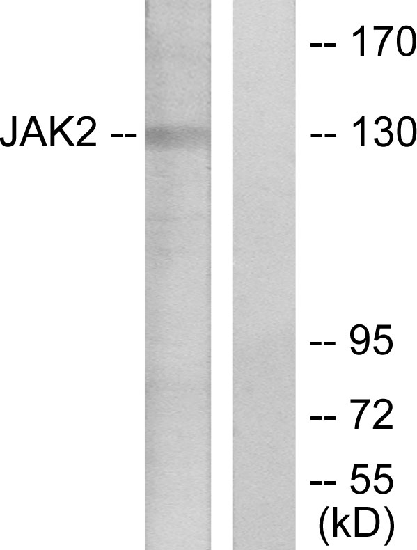
Figure 1. Western blot analysis of JAK2 using anti-JAK2 antibody (M00027). Electrophoresis was performed on a 5-20% SDS-PAGE gel at 70V (Stacking gel) / 90V (Resolving gel) for 2-3 hours. The sample well of each lane was loaded with 30 ug of sample under reducing conditions. Lane 1: human HepG2 whole cell lysates, Lane 2: human THP-1 whole cell lysates, Lane 3: rat NRK whole cell lysates, Lane 4: rat PC-12 whole cell lysates, Lane 5: mouse RAW264.7 whole cell lysates. After electrophoresis, proteins were transferred to a nitrocellulose membrane at 150 mA for 50-90 minutes. Blocked the membrane with 5% non-fat milk/TBS for 1.5 hour at RT. The membrane was incubated with rabbit anti-JAK2 antigen affinity purified monoclonal antibody (Catalog # M00027) at 1:500 overnight at 4°C, then washed with TBS-0.1%Tween 3 times with 5 minutes each and probed with a goat anti-rabbit IgG-HRP secondary antibody at a dilution of 1:1000 for 1.5 hour at RT. The signal is developed using an Enhanced Chemiluminescent detection (ECL) kit (Catalog # EK1002) with Tanon 5200 system. A specific band was detected for JAK2 at approximately 131 kDa. The expected band size for JAK2 is at 131 kDa.
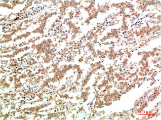
. JAK2 was detected in a paraffin-embedded section of human prostate cancer tissue. Heat mediated antigen retrieval was performed in EDTA buffer (pH 8.0, epitope retrieval solution). The tissue section was blocked with 10% goat serum. The tissue section was then incubated with 1:50 rabbit anti-JAK2 Antibody (M00027) overnight at 4°C. Peroxidase Conjugated Goat Anti-rabbit IgG was used as secondary antibody and incubated for 30 minutes at 37°C. The tissue section was developed using HRP Conjugated Rabbit IgG Super Vision Assay Kit (Catalog # SV0002) with DAB as the chromogen.")
 and anti-Beta Tubulin antibody (M01857-3). JAK2 was detected in immunocytochemical section of A549 cell. Enzyme antigen retrieval was performed using IHC enzyme antigen retrieval reagent (AR0022) for 15 mins. The cells were blocked with 10% goat serum. And then incubated at 1:50 with rabbit anti-JAK2 Antibody (M00027) and mouse anti-Beta Tubulin antibody (M01857-3) overnight at 4°C. DyLight?488 Conjugated Goat Anti-Rabbit IgG (BA1127) and Cy3 Conjugated Goat Anti-Mouse IgG (BA1031) were used as secondary antibody at 1:500 dilution and incubated for 30 minutes at 37°C. Visualize using a fluorescence microscope and filter sets appropriate for the label used.")
Figure 1. Western blot analysis of JAK2 using anti-JAK2 antibody (M00027). Electrophoresis was performed on a 5-20% SDS-PAGE gel at 70V (Stacking gel) / 90V (Resolving gel) for 2-3 hours. The sample well of each lane was loaded with 30 ug of sample under reducing conditions. Lane 1: human HepG2 whole cell lysates, Lane 2: human THP-1 whole cell lysates, Lane 3: rat NRK whole cell lysates, Lane 4: rat PC-12 whole cell lysates, Lane 5: mouse RAW264.7 whole cell lysates. After electrophoresis, proteins were transferred to a nitrocellulose membrane at 150 mA for 50-90 minutes. Blocked the membrane with 5% non-fat milk/TBS for 1.5 hour at RT. The membrane was incubated with rabbit anti-JAK2 antigen affinity purified monoclonal antibody (Catalog # M00027) at 1:500 overnight at 4°C, then washed with TBS-0.1%Tween 3 times with 5 minutes each and probed with a goat anti-rabbit IgG-HRP secondary antibody at a dilution of 1:1000 for 1.5 hour at RT. The signal is developed using an Enhanced Chemiluminescent detection (ECL) kit (Catalog # EK1002) with Tanon 5200 system. A specific band was detected for JAK2 at approximately 131 kDa. The expected band size for JAK2 is at 131 kDa.
Anti-JAK2 Monoclonal Antibody
M00027
ApplicationsImmunoFluorescence, ImmunoPrecipitation, Western Blot, ImmunoCytoChemistry, ImmunoHistoChemistry
Product group Antibodies
ReactivityHuman, Mouse, Rat
TargetJAK2
Overview
- SupplierBoster Bio
- Product NameAnti-JAK2 Monoclonal Antibody
- Delivery Days Customer9
- ApplicationsImmunoFluorescence, ImmunoPrecipitation, Western Blot, ImmunoCytoChemistry, ImmunoHistoChemistry
- CertificationResearch Use Only
- ClonalityMonoclonal
- Clone IDCOG-10
- Gene ID3717
- Target nameJAK2
- Target descriptionJanus kinase 2
- Target synonymsJTK10, tyrosine-protein kinase JAK2, JAK-2, Janus kinase 2 (a protein tyrosine kinase)
- HostRabbit
- IsotypeIgG
- Protein IDO60674
- Protein NameTyrosine-protein kinase JAK2
- Scientific DescriptionBoster Bio Anti-JAK2 Monoclonal Antibody catalog # M00027. Tested in WB, IHC, ICC/IF, IP applications. This antibody reacts with Human, Mouse, Rat.
- ReactivityHuman, Mouse, Rat
- Storage Instruction-20°C
- UNSPSC12352203
References
- Deng Z, Chen G, Shi Y, et al. Curcumin and its nano-formulations: Defining triple-negative breast cancer targets through network pharmacology, molecular docking, and experimental verification. Front Pharmacol. 2022,13:920514. doi: 10.3389/fphar.2022.920514Read this paper
- He J, Zhang W, Di T, et al. Water extract of sporoderm-broken spores of Ganoderma lucidum enhanced pd-l1 antibody efficiency through downregulation and relieved complications of pd-l1 monoclonal antibody. Biomed Pharmacother. 2020,131:110541. doi: 10.1016/j.biopha.2020.110541Read this paper
- Xu CP, Sun HT, Yang YJ, et al. ELP2 negatively regulates osteoblastic differentiation impaired by tumor necrosis factor α in MC3T3-E1 cells through STAT3 activation. J Cell Physiol. 2019,234(10):18075-18085. doi: 10.1002/jcp.28440Read this paper
- Liu K, Gao H, Wang Q, et al. Hispidulin suppresses cell growth and metastasis by targeting PIM1 through JAK2/STAT3 signaling in colorectal cancer. Cancer Sci. 2018,109(5):1369-1381. doi: 10.1111/cas.13575Read this paper
Datasheet
MSDS
Related products
Product group Antibodies
Anti-JAK2 AntibodyA96171
ApplicationsWestern Blot, ELISA, ImmunoHistoChemistry
ReactivityHuman, Mouse, Rat
- SizePrice

Product group Antibodies
Anti-JAK2 AntibodyHPA040820
ApplicationsImmunoHistoChemistry
ReactivityHuman
TargetJAK2
- SizePrice
Product group Antibodies
JAK2 Monoclonal AntibodyCSB-MA181103
ApplicationsELISA, ImmunoHistoChemistry
ReactivityHuman, Mouse, Rat
TargetJAK2
- SizePrice

Product group Antibodies
ApplicationsImmunoFluorescence, Western Blot, ImmunoCytoChemistry, ImmunoHistoChemistry
ReactivityPorcine
TargetJAK2
- SizePrice
Product group Antibodies
References
JAK2 Polyclonal AntibodyBS-0908R
ApplicationsFlow Cytometry, ImmunoFluorescence, Western Blot, ELISA, ImmunoCytoChemistry, ImmunoHistoChemistry, ImmunoHistoChemistry Frozen, ImmunoHistoChemistry Paraffin
ReactivityBovine, Canine, Chicken, Human, Mouse, Porcine, Rat
TargetJAK2
- SizePrice
Product group Antibodies
JAK2 (phospho Tyr1007/1008) antibodyGTX132784
ApplicationsWestern Blot
ReactivityHuman, Mouse
TargetJAK2
- SizePrice