


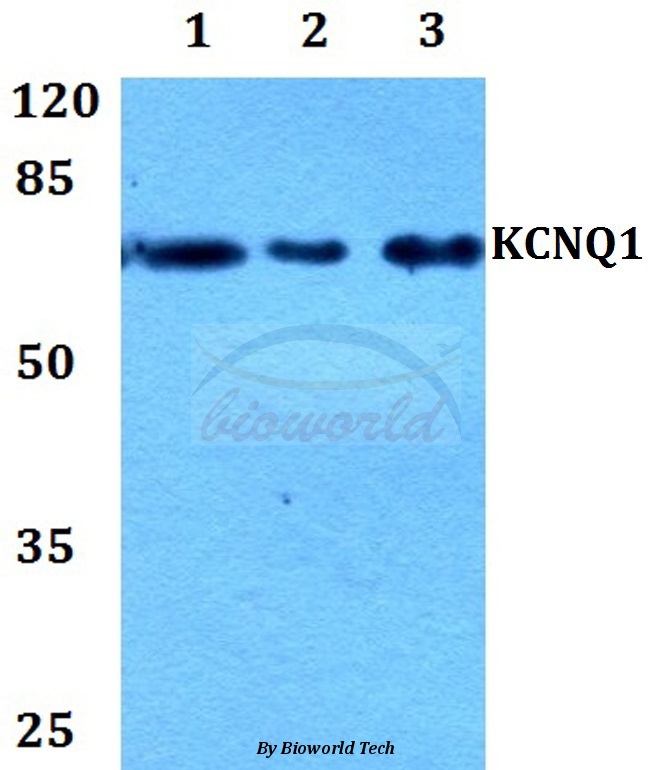
Figure 1. Western blot analysis of KCNQ1 using anti-KCNQ1 antibody (A00310-1). Electrophoresis was performed on a 5-20% SDS-PAGE gel at 70V (Stacking gel) / 90V (Resolving gel) for 2-3 hours. The sample well of each lane was loaded with 30 ug of sample under reducing conditions. Lane 1: human THP-1 whole cell lysates, Lane 2: rat stomach tissue lysates, Lane 3: rat lung tissue lysates, Lane 4: rat PC-12 whole cell lysates, Lane 5: mouse stomach tissue lysates, Lane 6: mouse lung tissue lysates, Lane 7: mouse NIH/3T3 whole cell lysates. After electrophoresis, proteins were transferred to a nitrocellulose membrane at 150 mA for 50-90 minutes. Blocked the membrane with 5% non-fat milk/TBS for 1.5 hour at RT. The membrane was incubated with rabbit anti-KCNQ1 antigen affinity purified polyclonal antibody (Catalog # A00310-1) at 0.5 microg/mL overnight at 4°C, then washed with TBS-0.1%Tween 3 times with 5 minutes each and probed with a goat anti-rabbit IgG-HRP secondary antibody at a dilution of 1:5000 for 1.5 hour at RT. The signal is developed using an Enhanced Chemiluminescent detection (ECL) kit (Catalog # EK1002) with Tanon 5200 system. A specific band was detected for KCNQ1 at approximately 75 kDa. The expected band size for KCNQ1 is at 75 kDa.
. KCNQ1 was detected in a paraffin-embedded section of human liver cancer tissue. Heat mediated antigen retrieval was performed in EDTA buffer (pH 8.0, epitope retrieval solution). The tissue section was blocked with 10% goat serum. The tissue section was then incubated with 2 microg/ml rabbit anti-KCNQ1 Antibody (A00310-1) overnight at 4°C. Peroxidase Conjugated Goat Anti-rabbit IgG was used as secondary antibody and incubated for 30 minutes at 37°C. The tissue section was developed using HRP Conjugated Rabbit IgG Super Vision Assay Kit (Catalog # SV0002) with DAB as the chromogen.")
. KCNQ1 was detected in a paraffin-embedded section of human lung cancer tissue. Heat mediated antigen retrieval was performed in EDTA buffer (pH 8.0, epitope retrieval solution). The tissue section was blocked with 10% goat serum. The tissue section was then incubated with 2 microg/ml rabbit anti-KCNQ1 Antibody (A00310-1) overnight at 4°C. Peroxidase Conjugated Goat Anti-rabbit IgG was used as secondary antibody and incubated for 30 minutes at 37°C. The tissue section was developed using HRP Conjugated Rabbit IgG Super Vision Assay Kit (Catalog # SV0002) with DAB as the chromogen.")
. KCNQ1 was detected in a paraffin-embedded section of human placenta tissue. Heat mediated antigen retrieval was performed in EDTA buffer (pH 8.0, epitope retrieval solution). The tissue section was blocked with 10% goat serum. The tissue section was then incubated with 2 microg/ml rabbit anti-KCNQ1 Antibody (A00310-1) overnight at 4°C. Peroxidase Conjugated Goat Anti-rabbit IgG was used as secondary antibody and incubated for 30 minutes at 37°C. The tissue section was developed using HRP Conjugated Rabbit IgG Super Vision Assay Kit (Catalog # SV0002) with DAB as the chromogen.")
. KCNQ1 was detected in a paraffin-embedded section of human breast cancer tissue. Heat mediated antigen retrieval was performed in EDTA buffer (pH 8.0, epitope retrieval solution). The tissue section was blocked with 10% goat serum. The tissue section was then incubated with 2 microg/ml rabbit anti-KCNQ1 Antibody (A00310-1) overnight at 4°C. Peroxidase Conjugated Goat Anti-rabbit IgG was used as secondary antibody and incubated for 30 minutes at 37°C. The tissue section was developed using HRP Conjugated Rabbit IgG Super Vision Assay Kit (Catalog # SV0002) with DAB as the chromogen.")
. KCNQ1 was detected in an immunocytochemical section of Hela cells. Enzyme antigen retrieval was performed using IHC enzyme antigen retrieval reagent (AR0022) for 15 mins. The cells were blocked with 10% goat serum. And then incubated with 5 microg/mL rabbit anti-KCNQ1 Antibody (A00310-1) overnight at 4°C. DyLight®488 Conjugated Goat Anti-Rabbit IgG (BA1127) was used as secondary antibody at 1:100 dilution and incubated for 30 minutes at 37°C. The section was counterstained with DAPI. Visualize using a fluorescence microscope and filter sets appropriate for the label used.")
. Overlay histogram showing U937 cells stained with A00310-1 (Blue line). To facilitate intracellular staining, cells were fixed with 4% paraformaldehyde and permeabilized with permeabilization buffer. The cells were blocked with 10% normal goat serum. And then incubated with rabbit anti-KCNQ1 Antibody (A00310-1, 1 microg/1x106 cells) for 30 min at 20°C. DyLight®488 conjugated goat anti-rabbit IgG (BA1127, 5-10 microg/1x106 cells) was used as secondary antibody for 30 minutes at 20°C. Isotype control antibody (Green line) was rabbit IgG (1 microg/1x106) used under the same conditions. Unlabelled sample without incubation with primary antibody and secondary antibody (Red line) was used as a blank control.")
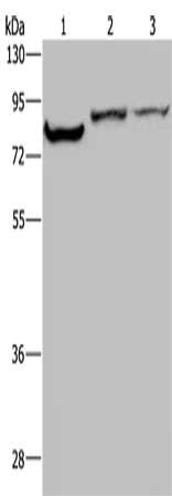
Figure 1. Western blot analysis of KCNQ1 using anti-KCNQ1 antibody (A00310-1). Electrophoresis was performed on a 5-20% SDS-PAGE gel at 70V (Stacking gel) / 90V (Resolving gel) for 2-3 hours. The sample well of each lane was loaded with 30 ug of sample under reducing conditions. Lane 1: human THP-1 whole cell lysates, Lane 2: rat stomach tissue lysates, Lane 3: rat lung tissue lysates, Lane 4: rat PC-12 whole cell lysates, Lane 5: mouse stomach tissue lysates, Lane 6: mouse lung tissue lysates, Lane 7: mouse NIH/3T3 whole cell lysates. After electrophoresis, proteins were transferred to a nitrocellulose membrane at 150 mA for 50-90 minutes. Blocked the membrane with 5% non-fat milk/TBS for 1.5 hour at RT. The membrane was incubated with rabbit anti-KCNQ1 antigen affinity purified polyclonal antibody (Catalog # A00310-1) at 0.5 microg/mL overnight at 4°C, then washed with TBS-0.1%Tween 3 times with 5 minutes each and probed with a goat anti-rabbit IgG-HRP secondary antibody at a dilution of 1:5000 for 1.5 hour at RT. The signal is developed using an Enhanced Chemiluminescent detection (ECL) kit (Catalog # EK1002) with Tanon 5200 system. A specific band was detected for KCNQ1 at approximately 75 kDa. The expected band size for KCNQ1 is at 75 kDa.
Anti-KCNQ1 Antibody Picoband(r)
A00310-1-CARRIER-FREE
ApplicationsFlow Cytometry, ImmunoFluorescence, Western Blot, ImmunoCytoChemistry, ImmunoHistoChemistry
Product group Antibodies
ReactivityHuman, Mouse, Rat
TargetKCNQ1
Overview
- SupplierBoster Bio
- Product NameAnti-KCNQ1 Antibody Picoband(r)
- Delivery Days Customer9
- ApplicationsFlow Cytometry, ImmunoFluorescence, Western Blot, ImmunoCytoChemistry, ImmunoHistoChemistry
- CertificationResearch Use Only
- ClonalityPolyclonal
- Concentration500 ug/ml
- Gene ID3784
- Target nameKCNQ1
- Target descriptionpotassium voltage-gated channel subfamily Q member 1
- Target synonymsATFB1, ATFB3, JLNS1, KCNA8, KCNA9, KVLQT1, Kv1.9, Kv7.1, LQT, LQT1, RWS, SQT2, WRS, potassium voltage-gated channel subfamily KQT member 1, IKs producing slow voltage-gated potassium channel subunit alpha KvLQT1, kidney and cardiac voltage dependend K+ channel, potassium channel, voltage gated KQT-like subfamily Q, member 1, potassium voltage-gated channel, KQT-like subfamily, member 1, slow delayed rectifier channel subunit, voltage-gated potassium channel subunit Kv7.1
- HostRabbit
- IsotypeIgG
- Protein IDP51787
- Protein NamePotassium voltage-gated channel subfamily KQT member 1
- Scientific DescriptionBoster Bio Anti-KCNQ1 Antibody Picoband® catalog # A00310-1. Tested in Flow Cytometry, IF, IHC, ICC, WB applications. This antibody reacts with Human, Mouse, Rat. The brand Picoband indicates this is a premium antibody that guarantees superior quality, high affinity, and strong signals with minimal background in Western blot applications. Only our best-performing antibodies are designated as Picoband, ensuring unmatched performance.
- ReactivityHuman, Mouse, Rat
- Storage Instruction-20°C,2°C to 8°C
- UNSPSC12352203
Related products
Product group Antibodies
Anti-Kv7.1 AntibodyA28399
ApplicationsWestern Blot
ReactivityHuman, Mouse, Rat
- SizePrice
Product group Antibodies
Anti-KCNQ1 Antibody144-02174
ApplicationsWestern Blot
ReactivityHuman, Mouse
TargetKCNQ1
- SizePrice
Product group Antibodies
References
KCNQ1 Polyclonal AntibodyBS-6760R
ApplicationsImmunoFluorescence, Western Blot, ELISA, ImmunoCytoChemistry, ImmunoHistoChemistry, ImmunoHistoChemistry Frozen, ImmunoHistoChemistry Paraffin
ReactivityBovine, Canine, Chicken, Human, Mouse, Rat
TargetKCNQ1
- SizePrice
Product group Antibodies

Goat anti-KCNQ1EB09214
ApplicationsWestern Blot, ELISA, ImmunoHistoChemistry
ReactivityBovine, Canine, Human, Mouse, Rat
TargetKCNQ1
- SizePrice
Product group Antibodies
KCNQ1 AntibodyCSB-PA237012
ApplicationsWestern Blot, ELISA
ReactivityHuman, Mouse, Rat
TargetKCNQ1
- SizePrice
Product group Antibodies

KCNQ1 / KVLQT1 AntibodyLS-C405102
ApplicationsWestern Blot, ELISA, ImmunoHistoChemistry
ReactivityHuman, Mouse, Rat
TargetKCNQ1
- SizePrice

Product group Antibodies
Anti-KCNQ1 AntibodyHPA048553
ApplicationsImmunoHistoChemistry
ReactivityHuman
TargetKCNQ1
- SizePrice
Product group Antibodies
KCNQ1 antibody [S37A-10]GTX41988
ApplicationsImmunoPrecipitation, Western Blot, ImmunoHistoChemistry
ReactivityHuman, Mouse, Rat
TargetKCNQ1
- SizePrice