Bio-Connect
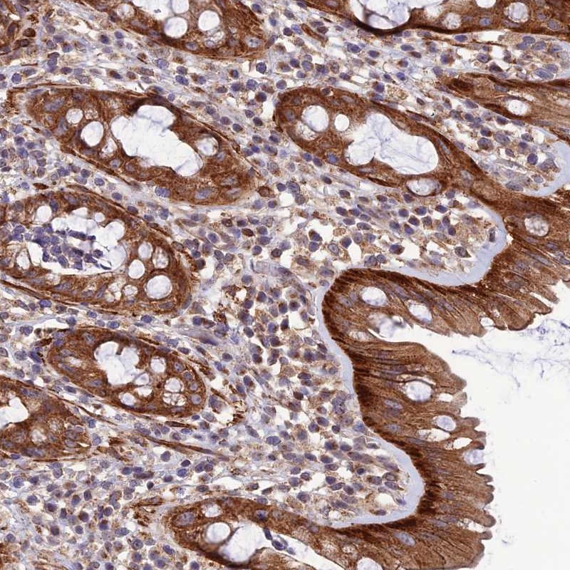
Immunohistochemical staining of human rectum shows strong cytoplasmic positivity in glandular cells.

Anti-LEKR1 Antibody

HPA044039
Atlas Antibodies
ApplicationsImmunoHistoChemistry
Product group Antibodies
TargetLEKR1
Sign in to order and to see your custom pricing.
Large volume orders?
Order with a bulk request

Overview

  • Supplier
    Atlas Antibodies
  • Product Name
    Anti-LEKR1
  • Delivery Days Customer
    4
  • Applications
    ImmunoHistoChemistry
  • Certification
    Research Use Only
  • Clonality
    Polyclonal
  • Conjugate
    Unconjugated
  • Gene ID389170
  • Target name
    LEKR1
  • Target description
    leucine, glutamate and lysine rich 1
  • Target synonyms
    leucine-, glutamate- and lysine-rich protein 1
  • Host
    Rabbit
  • Isotype
    IgG
  • Protein IDQ6ZMV7
  • Protein Name
    Leucine-, glutamate- and lysine-rich protein 1
  • Scientific Description
    Recombinant Protein Epitope Signature Tag (PrEST) antigen sequence
  • Storage Instruction
    -20°C,2°C to 8°C
  • UNSPSC
    12352203

Related products

Atlas Antibodies
Protein IDQ6ZMV7
  • SizePrice
Product group Antibodies
LifeSpan BioSciences
LEKR1 AntibodyLS-C820511
ApplicationsImmunoHistoChemistry, ImmunoHistoChemistry Paraffin
TargetLEKR1
  • SizePrice
Atlas Antibodies
Protein IDQ6ZMV7
  • SizePrice
Immunofluorescent staining of human cell line U-2 OS shows localization to nucleoplasm.
Product group Antibodies
Atlas Antibodies
ApplicationsImmunoCytoChemistry
TargetLEKR1
  • SizePrice
Figure 1. Western blot analysis of LEKR1 using anti-LEKR1 antibody (A15972-1). Electrophoresis was performed on a 5-20% SDS-PAGE gel at 70V (Stacking gel) / 90V (Resolving gel) for 2-3 hours. The sample well of each lane was loaded with 30 ug of sample under reducing conditions. Lane 1: human A431 whole cell lysates, Lane 2: human THP-1 whole cell lysates, Lane 3: human MCF-7 whole cell lysates, Lane 4: human SiHa whole cell lysates, Lane 5: rat C6 whole cell lysates, Lane 6: mouse NIH/3T3 whole cell lysates. After electrophoresis, proteins were transferred to a nitrocellulose membrane at 150 mA for 50-90 minutes. Blocked the membrane with 5% non-fat milk/TBS for 1.5 hour at RT. The membrane was incubated with rabbit anti-LEKR1 antigen affinity purified polyclonal antibody (Catalog # A15972-1) at 0.25 microg/mL overnight at 4°C, then washed with TBS-0.1%Tween 3 times with 5 minutes each and probed with a goat anti-rabbit IgG-HRP secondary antibody at a dilution of 1:5000 for 1.5 hour at RT. The signal is developed using an Enhanced Chemiluminescent detection (ECL) kit (Catalog # EK1002) with Tanon 5200 system. A specific band was detected for LEKR1 at approximately 16 kDa. The expected band size for LEKR1 is at 81 kDa.
Product group Antibodies
Boster Bio
Anti-LEKR1 Antibody Picoband(r)A15972-1-CARRIER-FREE
ApplicationsImmunoFluorescence, Western Blot, ELISA, ImmunoCytoChemistry, ImmunoHistoChemistry
TargetLEKR1
  • SizePrice