


Immunohistochemical staining of human rectum shows moderate cytoplasmic positivity in glandular cells.

Immunohistochemical staining of human rectum shows moderate cytoplasmic positivity in glandular cells.
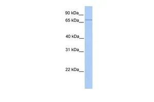
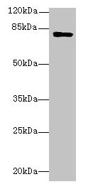
Anti-LMF2 Antibody
HPA062626
ApplicationsImmunoCytoChemistry, ImmunoHistoChemistry
Product group Antibodies
ReactivityHuman
TargetLMF2
Overview
- SupplierAtlas Antibodies
- Product NameAnti-LMF2 Antibody
- Delivery Days Customer4
- ApplicationsImmunoCytoChemistry, ImmunoHistoChemistry
- CertificationResearch Use Only
- ClonalityPolyclonal
- ConjugateUnconjugated
- Gene ID91289
- Target nameLMF2
- Target descriptionlipase maturation factor 2
- Target synonymsTMEM112B, TMEM153, lipase maturation factor 2, transmembrane protein 112B, transmembrane protein 153
- HostRabbit
- IsotypeIgG
- Protein IDQ9BU23
- Protein NameLipase maturation factor 2
- Scientific DescriptionRecombinant Protein Epitope Signature Tag (PrEST) antigen sequence
- ReactivityHuman
- Storage Instruction-20°C,2°C to 8°C
- UNSPSC41116161
Datasheet
MSDS
Related products
Product group Antibodies
LMF2 Polyclonal AntibodyCAC13803
ApplicationsImmunoFluorescence, Western Blot, ELISA
ReactivityMouse
TargetLMF2
- SizePrice
Product group Antibodies
Anti-LMF2 (C-term) Antibody102-20663
ApplicationsFlow Cytometry, Western Blot
TargetLMF2
- SizePrice
Product group Antibodies

TMEM153 / LMF2 AntibodyLS-C395972
ApplicationsWestern Blot, ELISA
ReactivityHuman, Mouse
TargetLMF2
- SizePrice
Product group Antibodies
LMF2 antibody, InternalGTX45908
ApplicationsWestern Blot
ReactivityHuman
TargetLMF2
- SizePrice
Product group Antibodies
LMF2 AntibodyCSB-PA013001LA01HU
ApplicationsImmunoFluorescence, Western Blot, ELISA
ReactivityHuman, Mouse
TargetLMF2
- SizePrice

Product group Antibodies
Anti-LMF2 Antibody Picoband(r)A13780-1-CARRIER-FREE
ApplicationsWestern Blot, ELISA
ReactivityHuman, Mouse, Rat
TargetLMF2
- SizePrice