


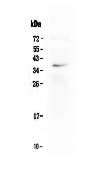
Figure 1. Western blot analysis of LOX 1/OLR1 using anti-LOX 1/OLR1 antibody (PA1833). Electrophoresis was performed on a 5-20% SDS-PAGE gel at 70V (Stacking gel) / 90V (Resolving gel) for 2-3 hours. The sample well of each lane was loaded with 30 ug of sample under reducing conditions. Lane 1: human prostatic carcinoma tissue (PCT) lysates, Lane 2: human cervical carcinoma tissue (CCAT) lysates, Lane 3: human gallbladder carcinoma tissue (GBCT) lysates, Lane 4: human intrahepatic cholangiocarcinom paracancerous tissue (ICCP) lysates. After electrophoresis, proteins were transferred to a nitrocellulose membrane at 150 mA for 50-90 minutes. Blocked the membrane with 5% non-fat milk/TBS for 1.5 hour at RT. The membrane was incubated with rabbit anti-LOX 1/OLR1 antigen affinity purified polyclonal antibody (Catalog # PA1833) at 0.5 microg/mL overnight at 4°C, then washed with TBS-0.1%Tween 3 times with 5 minutes each and probed with a goat anti-rabbit IgG-HRP secondary antibody at a dilution of 1:5000 for 1.5 hour at RT. The signal is developed using an Enhanced Chemiluminescent detection (ECL) kit (Catalog # EK1002) with Tanon 5200 system. A specific band was detected for LOX 1/OLR1 at approximately 48-52 kDa. The expected band size for LOX 1/OLR1 is at 31 kDa.
Figure 1. Western blot analysis of LOX 1/OLR1 using anti-LOX 1/OLR1 antibody (PA1833). Electrophoresis was performed on a 5-20% SDS-PAGE gel at 70V (Stacking gel) / 90V (Resolving gel) for 2-3 hours. The sample well of each lane was loaded with 30 ug of sample under reducing conditions. Lane 1: human prostatic carcinoma tissue (PCT) lysates, Lane 2: human cervical carcinoma tissue (CCAT) lysates, Lane 3: human gallbladder carcinoma tissue (GBCT) lysates, Lane 4: human intrahepatic cholangiocarcinom paracancerous tissue (ICCP) lysates. After electrophoresis, proteins were transferred to a nitrocellulose membrane at 150 mA for 50-90 minutes. Blocked the membrane with 5% non-fat milk/TBS for 1.5 hour at RT. The membrane was incubated with rabbit anti-LOX 1/OLR1 antigen affinity purified polyclonal antibody (Catalog # PA1833) at 0.5 microg/mL overnight at 4°C, then washed with TBS-0.1%Tween 3 times with 5 minutes each and probed with a goat anti-rabbit IgG-HRP secondary antibody at a dilution of 1:5000 for 1.5 hour at RT. The signal is developed using an Enhanced Chemiluminescent detection (ECL) kit (Catalog # EK1002) with Tanon 5200 system. A specific band was detected for LOX 1/OLR1 at approximately 48-52 kDa. The expected band size for LOX 1/OLR1 is at 31 kDa.
Anti-LOX 1/OLR1 Antibody Picoband(r)
PA1833
ApplicationsWestern Blot
Product group Antibodies
ReactivityHuman
TargetOLR1
Overview
- SupplierBoster Bio
- Product NameAnti-LOX 1/OLR1 Antibody Picoband(r)
- Delivery Days Customer9
- Application Supplier NoteTested Species: In-house tested species with positive results. Predicted Species: Species predicted to be fit for the product based on sequence similarities. Other applications have not been tested. Optimal dilutions should be determined by end users.
- ApplicationsWestern Blot
- Applications SupplierWB
- CertificationResearch Use Only
- ClonalityPolyclonal
- Concentration500 ug/ml
- Gene ID4973
- Target nameOLR1
- Target descriptionoxidized low density lipoprotein receptor 1
- Target synonymsCLEC8A, LOX1, LOXIN, SCARE1, SLOX1, oxidized low-density lipoprotein receptor 1, C-type lectin domain family 8 member A, hLOX-1, lectin-type oxidized LDL receptor 1, ox LDL receptor 1, oxidized low density lipoprotein (lectin-like) receptor 1, oxidized low-density lipoprotein receptor 1, soluble form, scavenger receptor class E, member 1
- HostRabbit
- IsotypeIgG
- Protein IDP78380
- Protein NameOxidized low-density lipoprotein receptor 1
- Scientific DescriptionBoster Bio Anti-LOX 1/OLR1 Antibody catalog # PA1833. Tested in WB applications. This antibody reacts with Human. The brand Picoband indicates this is a premium antibody that guarantees superior quality, high affinity, and strong signals with minimal background in Western blot applications. Only our best-performing antibodies are designated as Picoband, ensuring unmatched performance.
- ReactivityHuman
- Reactivity SupplierHuman, Rat
- Storage Instruction-20°C,2°C to 8°C
- UNSPSC12352203
Datasheet
MSDS
Related products
Product group Antibodies
OLR1 AntibodyCSB-PA016331LA01HU
ApplicationsImmunoFluorescence, Western Blot, ELISA, ImmunoHistoChemistry
ReactivityHuman, Rat
TargetOLR1
- SizePrice
Product group Antibodies
Anti-LOX-1/OLR1 Antibody Picoband(r)A00760-1-CARRIER-FREE
ApplicationsWestern Blot
ReactivityHuman
TargetOLR1
- SizePrice
Product group Antibodies

Anti-Lox-1 [AbAb01LOX1]AB03429-10.0-BT
ApplicationsELISA
ReactivityHuman
TargetOLR1
- SizePrice
Product group Antibodies
Anti-OLR1 AntibodyA30489
ApplicationsWestern Blot, ImmunoHistoChemistry
ReactivityHuman
- SizePrice
Product group Antibodies

ApplicationsWestern Blot, ELISA
ReactivityHuman
TargetOLR1
- SizePrice

Product group Antibodies
Anti-OLR1 AntibodyHPA050798
ApplicationsImmunoCytoChemistry
ReactivityHuman
TargetOLR1
- SizePrice
Product group Antibodies
OLR1 Polyclonal AntibodyCAC13853
ApplicationsImmunoFluorescence, Western Blot, ELISA, ImmunoHistoChemistry
ReactivityRat
TargetOLR1
- SizePrice
Product group Antibodies
OLR1 / LOX-1 antibody [B019M]GTX39124
ApplicationsWestern Blot, ELISA
ReactivityHuman
TargetOLR1
- SizePrice
Product group Antibodies
Anti-Lox1 (m) Antibody130-10587
ApplicationsELISA
ReactivityHuman
TargetOLR1
- SizePrice