

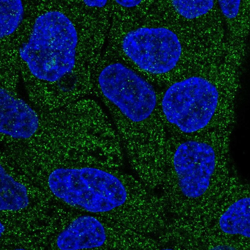
Immunofluorescent staining of human cell line RT4 shows localization to cytosol.
![Lane 1: Marker [kDa] 230, 130, 95, 72, 56, 36, 28, 17, 11. Lane 2: Human cell line RT-4. Lane 3: Human cell line U-251MG sp. Lane 4: Human plasma (IgG/HSA depleted). Lane 5: Human liver tissue](https://atlasantibodies.s3.amazonaws.com/images/wb/hpa031604-wb-1.jpg "Lane 1: Marker [kDa] 230, 130, 95, 72, 56, 36, 28, 17, 11. Lane 2: Human cell line RT-4. Lane 3: Human cell line U-251MG sp. Lane 4: Human plasma (IgG/HSA depleted). Lane 5: Human liver tissue")
Immunofluorescent staining of human cell line RT4 shows localization to cytosol.
Anti-LRRC1 Antibody
HPA031604
ApplicationsWestern Blot, ImmunoCytoChemistry
Product group Antibodies
ReactivityHuman
TargetLRRC1
Overview
- SupplierAtlas Antibodies
- Product NameAnti-LRRC1 Antibody
- Delivery Days Customer4
- ApplicationsWestern Blot, ImmunoCytoChemistry
- CertificationResearch Use Only
- ClonalityPolyclonal
- ConjugateUnconjugated
- Gene ID55227
- Target nameLRRC1
- Target descriptionleucine rich repeat containing 1
- Target synonymsLANO, dJ523E19.1, leucine-rich repeat-containing protein 1, LANO adapter protein, LAP (leucine-rich repeats and PDZ) and no PDZ protein, LAP and no PDZ protein
- HostRabbit
- IsotypeIgG
- Protein IDQ9BTT6
- Protein NameLeucine-rich repeat-containing protein 1
- Scientific DescriptionRecombinant Protein Epitope Signature Tag (PrEST) antigen sequence
- ReactivityHuman
- Storage Instruction-20°C,2°C to 8°C
- UNSPSC41116161
Datasheet
MSDS
Related products

Product group Antibodies
Anti-LRRC1 Antibody Picoband(r)A13956-2-CARRIER-FREE
ApplicationsWestern Blot, ELISA
ReactivityHuman, Mouse, Rat
TargetLRRC1
- SizePrice
Product group Antibodies
LRRC1 AntibodyCSB-PA013106GA01HU
ApplicationsELISA, ImmunoHistoChemistry
ReactivityHuman, Mouse, Rat
TargetLRRC1
- SizePrice
Product group Antibodies
Anti-LRRC1 AntibodyHPA031603
ApplicationsWestern Blot, ImmunoHistoChemistry
ReactivityHuman, Rat
TargetLRRC1
- SizePrice
Product group Antibodies
LRRC1 antibodyGTX119959
ApplicationsWestern Blot
ReactivityHuman
TargetLRRC1
- SizePrice
Product group Antibodies

LANO / LRRC1 AntibodyLS-C754176
ApplicationsELISA, ImmunoHistoChemistry
ReactivityHuman
TargetLRRC1
- SizePrice