

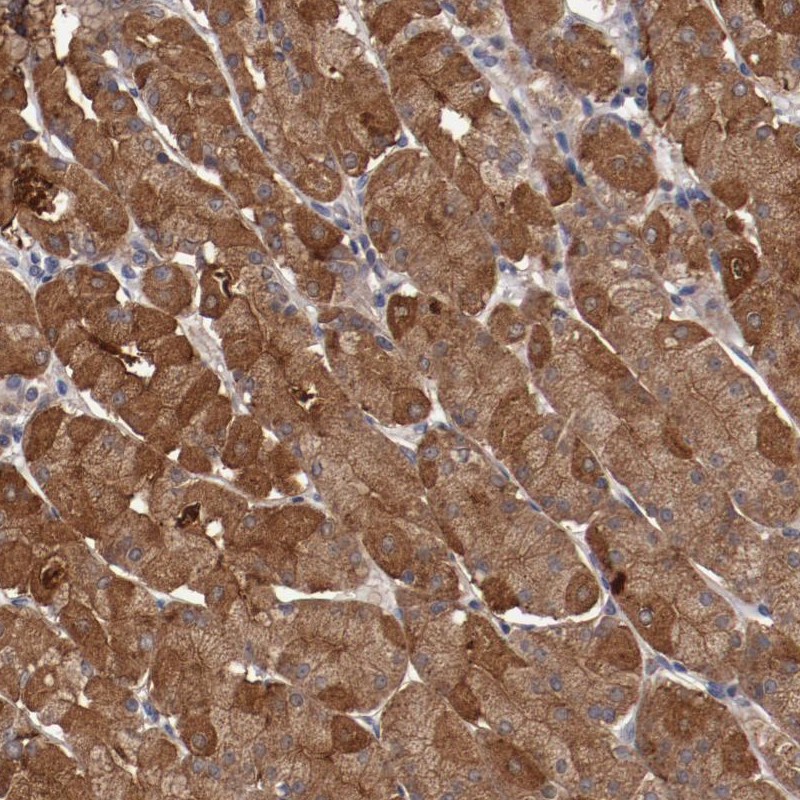
Immunohistochemical staining of human stomach shows cytoplasmic positivity in glandular cells.
Immunohistochemical staining of human stomach shows cytoplasmic positivity in glandular cells.
Anti-LRRC75B Antibody
HPA002540
ApplicationsImmunoHistoChemistry
Product group Antibodies
ReactivityHuman
TargetLRRC75B
Overview
- SupplierAtlas Antibodies
- Product NameAnti-LRRC75B Antibody
- Delivery Days Customer4
- ApplicationsImmunoHistoChemistry
- CertificationResearch Use Only
- ClonalityPolyclonal
- ConjugateUnconjugated
- Gene ID388886
- Target nameLRRC75B
- Target descriptionleucine rich repeat containing 75B
- Target synonymsC22orf36, FAM211B, leucine-rich repeat-containing protein 75B, family with sequence similarity 211, member B, leucine-rich repeat-containing protein C22orf36, leucine-rich repeat-containing protein FAM211B
- HostRabbit
- IsotypeIgG
- Protein IDQ2VPJ9
- Protein NameLeucine-rich repeat-containing protein 75B
- Scientific DescriptionRecombinant Protein Epitope Signature Tag (PrEST) antigen sequence
- ReactivityHuman
- Storage Instruction-20°C,2°C to 8°C
- UNSPSC41116161
Datasheet
MSDS
Related products

Product group Antibodies
Anti-LRRC75B Antibody Picoband(r)A18683-1-CARRIER-FREE
ApplicationsFlow Cytometry, Western Blot, ELISA, ImmunoHistoChemistry
ReactivityHuman, Mouse, Rat
TargetLRRC75B
- SizePrice
Product group Antibodies

LRRC75B / FAM211B Antibody (aa107-156)LS-C117187
ApplicationsWestern Blot
ReactivityHuman
TargetLRRC75B
- SizePrice