


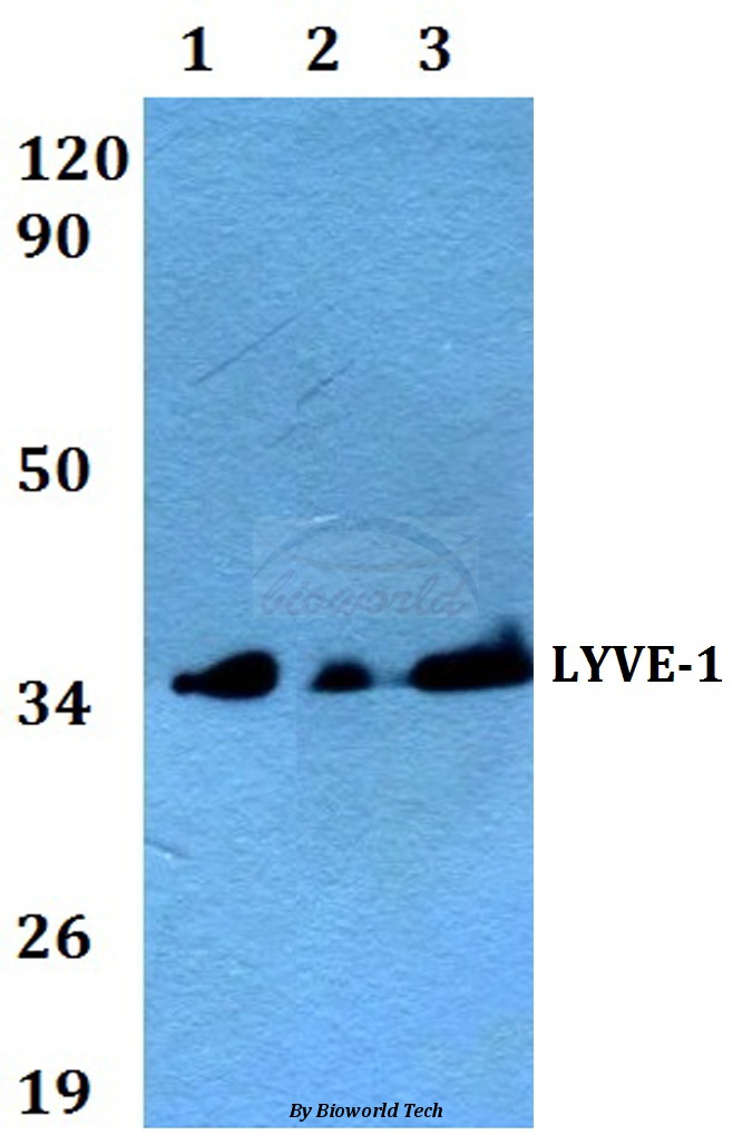
Figure 1. Western blot analysis of LYVE1 using anti-LYVE1 antibody (M04027). Electrophoresis was performed on a 5-20% SDS-PAGE gel at 70V (Stacking gel) / 90V (Resolving gel) for 2-3 hours. The sample well of each lane was loaded with 30 ug of sample under reducing conditions. Lane 1: human A549 whole cell lysates, Lane 2: human K562 whole cell lysates, Lane 3: human HepG2 whole cell lysates, Lane 4: human HEL whole cell lysates, Lane 5: rat C6 whole cell lysates, Lane 6: rat PC-12 whole cell lysates, Lane 7: mouse Neuro-2a whole cell lysates, Lane 8: mouse RAW264.7 whole cell lysates. After electrophoresis, proteins were transferred to a nitrocellulose membrane at 150 mA for 50-90 minutes. Blocked the membrane with 5% non-fat milk/TBS for 1.5 hour at RT. The membrane was incubated with rabbit anti-LYVE1 antigen affinity purified monoclonal antibody (Catalog # M04027) at 1:500 overnight at 4°C, then washed with TBS-0.1%Tween 3 times with 5 minutes each and probed with a goat anti-rabbit IgG-HRP secondary antibody at a dilution of 1:500 for 1.5 hour at RT. The signal is developed using an Enhanced Chemiluminescent detection (ECL) kit (Catalog # EK1002) with Tanon 5200 system. A specific band was detected for LYVE1 at approximately 29 kDa. The expected band size for LYVE1 is at 35 kDa.
Figure 1. Western blot analysis of LYVE1 using anti-LYVE1 antibody (M04027). Electrophoresis was performed on a 5-20% SDS-PAGE gel at 70V (Stacking gel) / 90V (Resolving gel) for 2-3 hours. The sample well of each lane was loaded with 30 ug of sample under reducing conditions. Lane 1: human A549 whole cell lysates, Lane 2: human K562 whole cell lysates, Lane 3: human HepG2 whole cell lysates, Lane 4: human HEL whole cell lysates, Lane 5: rat C6 whole cell lysates, Lane 6: rat PC-12 whole cell lysates, Lane 7: mouse Neuro-2a whole cell lysates, Lane 8: mouse RAW264.7 whole cell lysates. After electrophoresis, proteins were transferred to a nitrocellulose membrane at 150 mA for 50-90 minutes. Blocked the membrane with 5% non-fat milk/TBS for 1.5 hour at RT. The membrane was incubated with rabbit anti-LYVE1 antigen affinity purified monoclonal antibody (Catalog # M04027) at 1:500 overnight at 4°C, then washed with TBS-0.1%Tween 3 times with 5 minutes each and probed with a goat anti-rabbit IgG-HRP secondary antibody at a dilution of 1:500 for 1.5 hour at RT. The signal is developed using an Enhanced Chemiluminescent detection (ECL) kit (Catalog # EK1002) with Tanon 5200 system. A specific band was detected for LYVE1 at approximately 29 kDa. The expected band size for LYVE1 is at 35 kDa.
Anti-LYVE1 Rabbit Monoclonal Antibody
M04027
ApplicationsWestern Blot
Product group Antibodies
ReactivityHuman, Mouse, Rat
TargetLYVE1
Overview
- SupplierBoster Bio
- Product NameAnti-LYVE1 Rabbit Monoclonal Antibody
- Delivery Days Customer9
- ApplicationsWestern Blot
- CertificationResearch Use Only
- ClonalityMonoclonal
- Clone IDAOCO-12
- Gene ID10894
- Target nameLYVE1
- Target descriptionlymphatic vessel endothelial hyaluronan receptor 1
- Target synonymsCRSBP-1, HAR, LYVE-1, XLKD1, lymphatic vessel endothelial hyaluronic acid receptor 1, cell surface retention sequence binding protein-1, extracellular link domain-containing 1, extracellular link domain-containing protein 1, hyaluronic acid receptor
- HostRabbit
- IsotypeIgG
- Protein IDQ9Y5Y7
- Protein NameLymphatic vessel endothelial hyaluronic acid receptor 1
- Scientific DescriptionBoster Bio Anti-LYVE1 Rabbit Monoclonal Antibody catalog # M04027. Tested in WB application. This antibody reacts with Human, Mouse, Rat.
- ReactivityHuman, Mouse, Rat
- Storage Instruction-20°C
- UNSPSC12352203
References
- Du LC, Chen XC, Wang D, et al. VEGF-D-induced draining lymphatic enlargement and tumor lymphangiogenesis promote lymph node metastasis in a xenograft model of ovarian carcinoma. Reprod Biol Endocrinol. 2014,12:14. doi: 10.1186/1477-7827-12-14Read this paper
- Han DH, Song HK, Lee SY, et al. Upregulation of hyaluronan and its binding receptors in an experimental model of chronic cyclosporine nephropathy. Nephrology (Carlton). 2010,15(2):216-24. doi: 10.1111/j.1440-1797.2009.01167.xRead this paper
- Ling S, Lin H, Xiang D, et al. Clinical and experimental research of corneal lymphangiogenesis after keratoplasty. Ophthalmologica. 2008,222(5):308-16. doi: 10.1159/000144030Read this paper
Datasheet
MSDS
Related products
Product group Antibodies
LYVE1 AntibodyCSB-PA561582
ApplicationsWestern Blot, ELISA, ImmunoHistoChemistry
ReactivityHuman
TargetLYVE1
- SizePrice
Product group Antibodies
Anti-LYVE-1 AntibodyA24854
ApplicationsWestern Blot
ReactivityHuman, Mouse, Rat
- SizePrice

Product group Antibodies
Anti-LYVE1 AntibodyHPA042953
ApplicationsWestern Blot, ImmunoHistoChemistry
ReactivityHuman
TargetLYVE1
- SizePrice
Product group Antibodies

Goat anti-LYVE1EB12652
ApplicationsWestern Blot, ELISA
ReactivityHuman
TargetLYVE1
- SizePrice
Product group Antibodies

LYVE1 AntibodyLS-C349294
ApplicationsWestern Blot, ImmunoHistoChemistry
ReactivityHuman, Mouse
TargetLYVE1
- SizePrice
Product group Antibodies
Lyve1 Polyclonal AntibodyCAC07962
ApplicationsImmunoFluorescence, Western Blot, ELISA, ImmunoHistoChemistry
ReactivityMouse
TargetLYVE1
- SizePrice

Product group Antibodies
anti-Lyve1 (human), pAbAG-25T-0100
ApplicationsFlow Cytometry, Western Blot, ImmunoHistoChemistry
ReactivityHuman
TargetLYVE1
- SizePrice
Product group Antibodies
LYVE1 Recombinant AntibodyBSM-52811R
ApplicationsWestern Blot
ReactivityHuman, Mouse, Rat
TargetLYVE1
- SizePrice
Product group Antibodies
LYVE1 antibodyGTX133081
ApplicationsWestern Blot
ReactivityHuman
TargetLYVE1
- SizePrice