


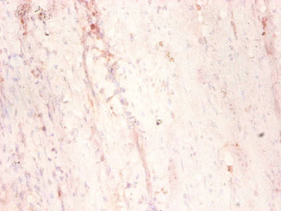
Immunohistochemical staining of human testis shows moderate cytoplasmic positivity in cells in seminiferous ducts.

Immunohistochemical staining of human testis shows moderate cytoplasmic positivity in cells in seminiferous ducts.
Anti-MAP1S Antibody
HPA050934
ApplicationsImmunoCytoChemistry, ImmunoHistoChemistry
Product group Antibodies
ReactivityHuman
TargetMAP1S
Overview
- SupplierAtlas Antibodies
- Product NameAnti-MAP1S Antibody
- Delivery Days Customer4
- ApplicationsImmunoCytoChemistry, ImmunoHistoChemistry
- CertificationResearch Use Only
- ClonalityPolyclonal
- ConjugateUnconjugated
- Gene ID55201
- Target nameMAP1S
- Target descriptionmicrotubule associated protein 1S
- Target synonymsBPY2IP1, C19orf5, MAP8, VCY2IP-1, VCY2IP1, microtubule-associated protein 1S, BPY2-interacting protein 1, MAP-1S, VCY2-interacting protein 1, microtubule-associated protein 8, variable charge Y chromosome 2-interacting protein 1
- HostRabbit
- IsotypeIgG
- Protein IDQ66K74
- Protein NameMicrotubule-associated protein 1S
- Scientific DescriptionRecombinant Protein Epitope Signature Tag (PrEST) antigen sequence
- ReactivityHuman
- Storage Instruction-20°C,2°C to 8°C
- UNSPSC41116161
Datasheet
MSDS
Related products

Product group Antibodies
Anti-MAP1S AntibodyHPA054637
ApplicationsImmunoCytoChemistry, ImmunoHistoChemistry
ReactivityHuman
TargetMAP1S
- SizePrice
Product group Antibodies

MAP1S Antibody (FITC)LS-C211386
ApplicationsWestern Blot, ELISA, ImmunoHistoChemistry, ImmunoHistoChemistry Paraffin
ReactivityHuman
TargetMAP1S
- SizePrice
Product group Antibodies
MAP1S AntibodyCSB-PA13549A0RB
ApplicationsELISA, ImmunoHistoChemistry
ReactivityHuman
TargetMAP1S
- SizePrice

Product group Antibodies
Anti-MAP1S Antibody Picoband(r)A04308-CARRIER-FREE
ApplicationsFlow Cytometry, Western Blot, ELISA, ImmunoHistoChemistry
ReactivityHuman, Mouse, Rat
TargetMAP1S
- SizePrice
Product group Antibodies
Map1S Polyclonal AntibodyCAC10418
ApplicationsELISA, ImmunoHistoChemistry
TargetMAP1S
- SizePrice