


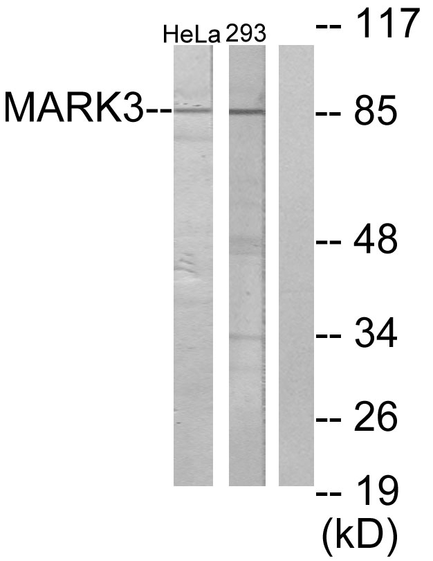
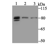
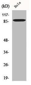
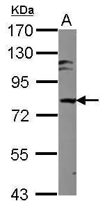
Figure 1. Western blot analysis of MARK3 using anti-MARK3 antibody (A05355-2). Electrophoresis was performed on a 5-20% SDS-PAGE gel at 70V (Stacking gel) / 90V (Resolving gel) for 2-3 hours. The sample well of each lane was loaded with 30 ug of sample under reducing conditions. Lane 1: human Hela whole cell lysates, Lane 2: human K562 whole cell lysates, Lane 3: rat NRK whole cell lysates, Lane 4: mouse EL-4 whole cell lysates, Lane 5: mouse 4T1 whole cell lysates, Lane 6: mouse J774A.1 whole cell lysates. After electrophoresis, proteins were transferred to a nitrocellulose membrane at 150 mA for 50-90 minutes. Blocked the membrane with 5% non-fat milk/TBS for 1.5 hour at RT. The membrane was incubated with rabbit anti-MARK3 antigen affinity purified polyclonal antibody (Catalog # A05355-2) at 0.5 microg/mL overnight at 4°C, then washed with TBS-0.1%Tween 3 times with 5 minutes each and probed with a goat anti-rabbit IgG-HRP secondary antibody at a dilution of 1:5000 for 1.5 hour at RT. The signal is developed using an Enhanced Chemiluminescent detection (ECL) kit (Catalog # EK1002) with Tanon 5200 system. A specific band was detected for MARK3 at approximately 80 kDa. The expected band size for MARK3 is at 80 kDa.
. MARK3 was detected in a paraffin-embedded section of human lung cancer tissue. Heat mediated antigen retrieval was performed in EDTA buffer (pH 8.0, epitope retrieval solution). The tissue section was blocked with 10% goat serum. The tissue section was then incubated with 2 microg/ml rabbit anti-MARK3 Antibody (A05355-2) overnight at 4°C. Peroxidase Conjugated Goat Anti-rabbit IgG was used as secondary antibody and incubated for 30 minutes at 37°C. The tissue section was developed using HRP Conjugated Rabbit IgG Super Vision Assay Kit (Catalog # SV0002) with DAB as the chromogen.")
. MARK3 was detected in a paraffin-embedded section of human thyroid cancer tissue. Heat mediated antigen retrieval was performed in EDTA buffer (pH 8.0, epitope retrieval solution). The tissue section was blocked with 10% goat serum. The tissue section was then incubated with 2 microg/ml rabbit anti-MARK3 Antibody (A05355-2) overnight at 4°C. Peroxidase Conjugated Goat Anti-rabbit IgG was used as secondary antibody and incubated for 30 minutes at 37°C. The tissue section was developed using HRP Conjugated Rabbit IgG Super Vision Assay Kit (Catalog # SV0002) with DAB as the chromogen.")
. MARK3 was detected in a paraffin-embedded section of mouse brain tissue. Heat mediated antigen retrieval was performed in EDTA buffer (pH 8.0, epitope retrieval solution). The tissue section was blocked with 10% goat serum. The tissue section was then incubated with 2 microg/ml rabbit anti-MARK3 Antibody (A05355-2) overnight at 4°C. Peroxidase Conjugated Goat Anti-rabbit IgG was used as secondary antibody and incubated for 30 minutes at 37°C. The tissue section was developed using HRP Conjugated Rabbit IgG Super Vision Assay Kit (Catalog # SV0002) with DAB as the chromogen.")
. MARK3 was detected in a paraffin-embedded section of rat brain tissue. Heat mediated antigen retrieval was performed in EDTA buffer (pH 8.0, epitope retrieval solution). The tissue section was blocked with 10% goat serum. The tissue section was then incubated with 2 microg/ml rabbit anti-MARK3 Antibody (A05355-2) overnight at 4°C. Peroxidase Conjugated Goat Anti-rabbit IgG was used as secondary antibody and incubated for 30 minutes at 37°C. The tissue section was developed using HRP Conjugated Rabbit IgG Super Vision Assay Kit (Catalog # SV0002) with DAB as the chromogen.")
. Overlay histogram showing 293T cells stained with A05355-2 (Blue line). To facilitate intracellular staining, cells were fixed with 4% paraformaldehyde and permeabilized with permeabilization buffer. The cells were blocked with 10% normal goat serum. And then incubated with rabbit anti-MARK3 Antibody (A05355-2, 1 microg/1x106 cells) for 30 min at 20°C. DyLight®488 conjugated goat anti-rabbit IgG (BA1127, 5-10 microg/1x106 cells) was used as secondary antibody for 30 minutes at 20°C. Isotype control antibody (Green line) was rabbit IgG (1 microg/1x106) used under the same conditions. Unlabelled sample (Red line) was also used as a control.")
Figure 1. Western blot analysis of MARK3 using anti-MARK3 antibody (A05355-2). Electrophoresis was performed on a 5-20% SDS-PAGE gel at 70V (Stacking gel) / 90V (Resolving gel) for 2-3 hours. The sample well of each lane was loaded with 30 ug of sample under reducing conditions. Lane 1: human Hela whole cell lysates, Lane 2: human K562 whole cell lysates, Lane 3: rat NRK whole cell lysates, Lane 4: mouse EL-4 whole cell lysates, Lane 5: mouse 4T1 whole cell lysates, Lane 6: mouse J774A.1 whole cell lysates. After electrophoresis, proteins were transferred to a nitrocellulose membrane at 150 mA for 50-90 minutes. Blocked the membrane with 5% non-fat milk/TBS for 1.5 hour at RT. The membrane was incubated with rabbit anti-MARK3 antigen affinity purified polyclonal antibody (Catalog # A05355-2) at 0.5 microg/mL overnight at 4°C, then washed with TBS-0.1%Tween 3 times with 5 minutes each and probed with a goat anti-rabbit IgG-HRP secondary antibody at a dilution of 1:5000 for 1.5 hour at RT. The signal is developed using an Enhanced Chemiluminescent detection (ECL) kit (Catalog # EK1002) with Tanon 5200 system. A specific band was detected for MARK3 at approximately 80 kDa. The expected band size for MARK3 is at 80 kDa.
Anti-MARK3 Antibody Picoband(r)
A05355-2-CARRIER-FREE
ApplicationsFlow Cytometry, Western Blot, ELISA, ImmunoHistoChemistry
Product group Antibodies
ReactivityHuman, Mouse, Rat
TargetMARK3
Overview
- SupplierBoster Bio
- Product NameAnti-MARK3 Antibody Picoband(r)
- Delivery Days Customer9
- ApplicationsFlow Cytometry, Western Blot, ELISA, ImmunoHistoChemistry
- CertificationResearch Use Only
- ClonalityPolyclonal
- Concentration500 ug/ml
- Gene ID4140
- Target nameMARK3
- Target descriptionmicrotubule affinity regulating kinase 3
- Target synonymsCTAK1, KP78, PAR1A, Par-1a, VIPB, MAP/microtubule affinity-regulating kinase 3, C-TAK1, ELKL motif kinase 2, EMK-2, cdc25C-associated protein kinase 1, protein kinase STK10, ser/Thr protein kinase PAR-1, serine/threonine-protein kinase p78
- HostRabbit
- IsotypeIgG
- Protein IDP27448
- Protein NameMAP/microtubule affinity-regulating kinase 3
- Scientific DescriptionBoster Bio Anti-MARK3 Antibody Picoband® catalog # A05355-2. Tested in ELISA, IHC, WB, Flow Cytometry applications. This antibody reacts with Human, Mouse, Rat. The brand Picoband indicates this is a premium antibody that guarantees superior quality, high affinity, and strong signals with minimal background in Western blot applications. Only our best-performing antibodies are designated as Picoband, ensuring unmatched performance.
- ReactivityHuman, Mouse, Rat
- Storage Instruction-20°C,2°C to 8°C
- UNSPSC12352203
Related products
Product group Antibodies
Anti-MARK3 AntibodyA95843
ApplicationsWestern Blot, ELISA, ImmunoHistoChemistry
ReactivityHuman, Mouse, Rat
- SizePrice
Product group Antibodies
Anti-Mark3 (C-term) Antibody102-21798
ApplicationsWestern Blot
TargetMARK3
- SizePrice
Product group Antibodies

C-TAK1 / MARK3 AntibodyLS-C830004
ApplicationsELISA, ImmunoHistoChemistry
ReactivityHuman, Mouse, Rat
TargetMARK3
- SizePrice
Product group Antibodies
MARK3 Recombinant AntibodyBSM-54462R
ApplicationsFlow Cytometry, ImmunoFluorescence, ImmunoPrecipitation, Western Blot, ImmunoCytoChemistry, ImmunoHistoChemistry, ImmunoHistoChemistry Paraffin
ReactivityHuman, Mouse, Rat
TargetMARK3
- SizePrice
Product group Antibodies
MARK3 AntibodyCSB-PA001817
ApplicationsImmunoFluorescence, Western Blot, ELISA, ImmunoHistoChemistry
ReactivityHuman, Mouse, Rat
TargetMARK3
- SizePrice
Product group Antibodies
MARK3 Polyclonal AntibodyCAC13108
ApplicationsImmunoFluorescence, ELISA, ImmunoHistoChemistry
TargetMARK3
- SizePrice

Product group Antibodies
Anti-MARK3 AntibodyHPA024652
ApplicationsWestern Blot, ImmunoHistoChemistry
ReactivityHuman, Mouse, Rat
TargetMARK3
- SizePrice
Product group Antibodies
Mark3 antibodyGTX111538
ApplicationsWestern Blot, ImmunoHistoChemistry, ImmunoHistoChemistry Paraffin
ReactivityHuman
TargetMARK3
- SizePrice