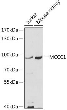
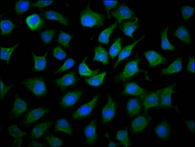
Bio-Connect
Anti-MCCC1 Antibody

Anti-MCCC1 Antibody

A11216
Antibodies.com
ApplicationsWestern Blot, ImmunoHistoChemistry
Product group Antibodies
Sign in to order and to see your custom pricing.
Large volume orders?
Order with a bulk request

Overview

  • Supplier
    Antibodies.com
  • Product Name
    Anti-MCCC1 Antibody
  • Delivery Days Customer
    7
  • Applications
    Western Blot, ImmunoHistoChemistry
  • Certification
    Research Use Only
  • Clonality
    Polyclonal
  • Conjugate
    Unconjugated
  • Host
    Rabbit
  • Isotype
    IgG
  • Scientific Description
    Rabbit polyclonal antibody to MCCC1.
  • UNSPSC
    12352203

Related products

Product group Antibodies
RayBiotech
ApplicationsWestern Blot, ImmunoHistoChemistry
TargetMCCC1
  • SizePrice
Immunofluorescence staining of A549 cells with CSB-PA853497EA01HU at 1:166, counter-stained with DAPI. The cells were fixed in 4% formaldehyde, permeabilized using 0.2% Triton X-100 and blocked in 10% normal Goat Serum. The cells were then incubated with the antibody overnight at 4°C. The secondary antibody was Alexa Fluor 488-congugated AffiniPure Goat Anti-Rabbit IgG(H+L).
Product group Antibodies
Cusabio
MCCC1 AntibodyCSB-PA853497EA01HU
ApplicationsImmunoFluorescence, ELISA
ReactivityHuman
TargetMCCC1
  • SizePrice
Product group Antibodies
LifeSpan BioSciences
MCCC1 AntibodyLS-C832139
ApplicationsWestern Blot, ELISA, ImmunoHistoChemistry
TargetMCCC1
  • SizePrice
Immunohistochemical staining of human cerebral cortex shows strong cytoplasmic positivity in neuronal cells.
Product group Antibodies
Atlas Antibodies
ApplicationsWestern Blot, ImmunoCytoChemistry, ImmunoHistoChemistry
ReactivityHuman
TargetMCCC1
  • SizePrice
Figure 1. Western blot analysis of MCCC1 using anti-MCCC1 antibody (A07441-1). Electrophoresis was performed on a 5-20% SDS-PAGE gel at 70V (Stacking gel) / 90V (Resolving gel) for 2-3 hours. The sample well of each lane was loaded with 30 ug of sample under reducing conditions. Lane 1: human 293T whole cell lysates, Lane 2: human SiHa whole cell lysates, Lane 3: human RT4 whole cell lysates, Lane 4: human Jurkat whole cell lysates, Lane 5: rat liver tissue lysates, Lane 6: rat heart tissue lysates, Lane 3: mouse liver tissue lysates, Lane 3: mouse heart tissue lysates. After electrophoresis, proteins were transferred to a nitrocellulose membrane at 150 mA for 50-90 minutes. Blocked the membrane with 5% non-fat milk/TBS for 1.5 hour at RT. The membrane was incubated with rabbit anti-MCCC1 antigen affinity purified polyclonal antibody (Catalog # A07441-1) at 0.5 microg/mL overnight at 4°C, then washed with TBS-0.1%Tween 3 times with 5 minutes each and probed with a goat anti-rabbit IgG-HRP secondary antibody at a dilution of 1:5000 for 1.5 hour at RT. The signal is developed using an Enhanced Chemiluminescent detection (ECL) kit (Catalog # EK1002) with Tanon 5200 system. A specific band was detected for MCCC1 at approximately 80 kDa. The expected band size for MCCC1 is at 80 kDa.
Product group Antibodies
Boster Bio
Anti-MCCC1 Antibody Picoband(r)A07441-1-CARRIER-FREE
ApplicationsImmunoFluorescence, Western Blot, ELISA, ImmunoCytoChemistry, ImmunoHistoChemistry
TargetMCCC1
  • SizePrice