


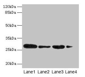
Figure 1. Western blot analysis of MED7 using anti-MED7 antibody (A09822). Electrophoresis was performed on a 5-20% SDS-PAGE gel at 70V (Stacking gel) / 90V (Resolving gel) for 2-3 hours. The sample well of each lane was loaded with 30 ug of sample under reducing conditions. Lane 1: human PC-3 whole cell lysates, Lane 2: human Caco-2 whole cell lysates, Lane 3: human A549 whole cell lysates, Lane 4: human Hela whole cell lysates, Lane 5: rat C6 whole cell lysates, Lane 6: rat PC-12 whole cell lysates, Lane 7: mouse RAW264.7 whole cell lysates, Lane 8: mouse NIH/3T3 whole cell lysates. After electrophoresis, proteins were transferred to a nitrocellulose membrane at 150 mA for 50-90 minutes. Blocked the membrane with 5% non-fat milk/TBS for 1.5 hour at RT. The membrane was incubated with rabbit anti-MED7 antigen affinity purified polyclonal antibody (Catalog # A09822) at 0.25 microg/mL overnight at 4°C, then washed with TBS-0.1%Tween 3 times with 5 minutes each and probed with a goat anti-rabbit IgG-HRP secondary antibody at a dilution of 1:5000 for 1.5 hour at RT. The signal is developed using an Enhanced Chemiluminescent detection (ECL) kit (Catalog # EK1002) with Tanon 5200 system. A specific band was detected for MED7 at approximately 27 kDa. The expected band size for MED7 is at 27 kDa.
 and anti-Beta Tubulin antibody (M01857-3). MED7 was detected in immunocytochemical section of A549 cell. Enzyme antigen retrieval was performed using IHC enzyme antigen retrieval reagent (AR0022) for 15 mins. The cells were blocked with 10% goat serum. And then incubated with 5 microg/mL rabbit anti-MED7 Antibody (A09822) and mouse anti-Beta Tubulin antibody (M01857-3) overnight at 4°C. DyLight®594 Conjugated Goat Anti-Rabbit IgG (BA1142) and DyLight®488 Conjugated Goat Anti-Mouse IgG (BA1126) were used as secondary antibody at 1:500 dilution and incubated for 30 minutes at 37°C. Visualize using a fluorescence microscope and filter sets appropriate for the label used.")
Figure 1. Western blot analysis of MED7 using anti-MED7 antibody (A09822). Electrophoresis was performed on a 5-20% SDS-PAGE gel at 70V (Stacking gel) / 90V (Resolving gel) for 2-3 hours. The sample well of each lane was loaded with 30 ug of sample under reducing conditions. Lane 1: human PC-3 whole cell lysates, Lane 2: human Caco-2 whole cell lysates, Lane 3: human A549 whole cell lysates, Lane 4: human Hela whole cell lysates, Lane 5: rat C6 whole cell lysates, Lane 6: rat PC-12 whole cell lysates, Lane 7: mouse RAW264.7 whole cell lysates, Lane 8: mouse NIH/3T3 whole cell lysates. After electrophoresis, proteins were transferred to a nitrocellulose membrane at 150 mA for 50-90 minutes. Blocked the membrane with 5% non-fat milk/TBS for 1.5 hour at RT. The membrane was incubated with rabbit anti-MED7 antigen affinity purified polyclonal antibody (Catalog # A09822) at 0.25 microg/mL overnight at 4°C, then washed with TBS-0.1%Tween 3 times with 5 minutes each and probed with a goat anti-rabbit IgG-HRP secondary antibody at a dilution of 1:5000 for 1.5 hour at RT. The signal is developed using an Enhanced Chemiluminescent detection (ECL) kit (Catalog # EK1002) with Tanon 5200 system. A specific band was detected for MED7 at approximately 27 kDa. The expected band size for MED7 is at 27 kDa.
Anti-MED7 Antibody Picoband(r)
A09822-CARRIER-FREE
ApplicationsImmunoFluorescence, Western Blot, ELISA, ImmunoCytoChemistry
Product group Antibodies
ReactivityHuman, Mouse, Rat
TargetMED7
Overview
- SupplierBoster Bio
- Product NameAnti-MED7 Antibody Picoband(r)
- Delivery Days Customer9
- ApplicationsImmunoFluorescence, Western Blot, ELISA, ImmunoCytoChemistry
- CertificationResearch Use Only
- ClonalityPolyclonal
- Concentration500 ug/ml
- Gene ID9443
- Target nameMED7
- Target descriptionmediator complex subunit 7
- Target synonymsARC34, CRSP33, CRSP9, mediator of RNA polymerase II transcription subunit 7, CRSP complex subunit 9, RNA polymerase transcriptional regulation mediator subunit 7 homolog, activator-recruited cofactor 34 kDa component, cofactor required for Sp1 transcriptional activation subunit 9, cofactor required for Sp1 transcriptional activation, subunit 9 (33kD), cofactor required for Sp1 transcriptional activation, subunit 9, 33kDa, transcriptional coactivator CRSP33
- HostRabbit
- Protein IDO43513
- Protein NameMediator of RNA polymerase II transcription subunit 7
- Scientific DescriptionBoster Bio Anti-MED7 Antibody Picoband® catalog # A09822. Tested in WB, ICC/IF, ELISA applications. This antibody reacts with Human, Mouse, Rat. The brand Picoband indicates this is a premium antibody that guarantees superior quality, high affinity, and strong signals with minimal background in Western blot applications. Only our best-performing antibodies are designated as Picoband, ensuring unmatched performance.
- ReactivityHuman, Mouse, Rat
- Storage Instruction-20°C,2°C to 8°C
- UNSPSC12352203
Related products
Product group Antibodies
Anti-MED7 Antibody144-64086
ApplicationsWestern Blot
ReactivityHuman, Mouse
TargetMED7
- SizePrice
Product group Antibodies
MED7 Polyclonal AntibodyCAC13822
ApplicationsWestern Blot, ELISA
ReactivityMouse
TargetMED7
- SizePrice
Product group Antibodies
CRSP9 Polyclonal AntibodyBS-11427R
ApplicationsImmunoFluorescence, Western Blot, ELISA, ImmunoCytoChemistry, ImmunoHistoChemistry, ImmunoHistoChemistry Frozen, ImmunoHistoChemistry Paraffin
ReactivityBovine, Canine, Chicken, Equine, Human, Mouse, Porcine, Rat, Sheep, Zebra Fish
TargetMED7
- SizePrice
Product group Antibodies
MED7 AntibodyCSB-PA013667LA01HU
ApplicationsWestern Blot, ELISA
ReactivityHuman, Mouse
TargetMED7
- SizePrice

Product group Antibodies
Anti-MED7 AntibodyHPA067181
ApplicationsImmunoCytoChemistry
ReactivityHuman
TargetMED7
- SizePrice
Product group Antibodies

MED7 / CRSP9 AntibodyLS-C376103
ApplicationsWestern Blot, ELISA
ReactivityHuman, Mouse
TargetMED7
- SizePrice
Product group Antibodies
CRSP9 antibody [C2C3], C-termGTX108522
ApplicationsWestern Blot
ReactivityHuman
TargetMED7
- SizePrice
Product group Antibodies
Anti-MED7 AntibodyA88999
ApplicationsWestern Blot
ReactivityHuman, Mouse
- SizePrice