


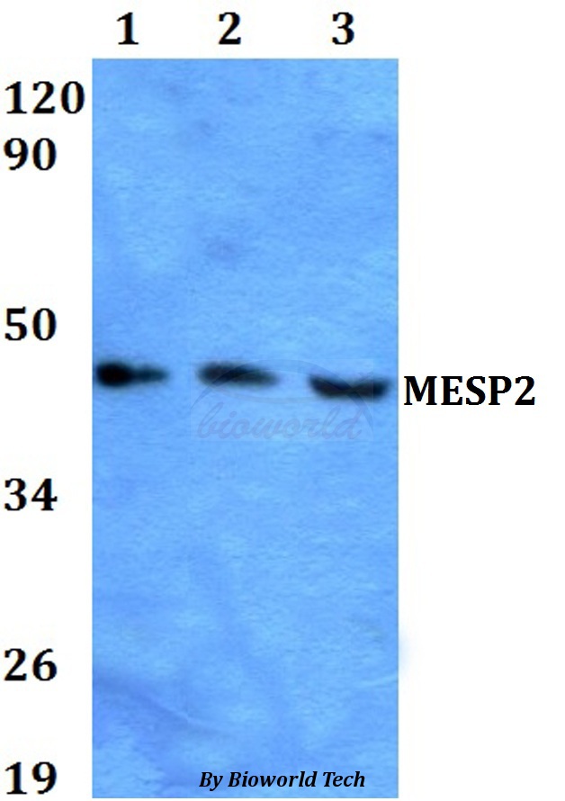
Figure 1. Western blot analysis of Mesp2 using anti-Mesp2 antibody (A07046-1). Electrophoresis was performed on a 5-20% SDS-PAGE gel at 70V (Stacking gel) / 90V (Resolving gel) for 2-3 hours. The sample well of each lane was loaded with 50ug of sample under reducing conditions. Lane 1: human placenta tissue lysates, Lane 2: human A549 whole cell lysates, Lane 3: human SW620 whole cell lysates. After Electrophoresis, proteins were transferred to a Nitrocellulose membrane at 150mA for 50-90 minutes. Blocked the membrane with 5% Non-fat Milk/ TBS for 1.5 hour at RT. The membrane was incubated with rabbit anti-Mesp2 antigen affinity purified polyclonal antibody (Catalog # A07046-1) at 0.5 microg/mL overnight at 4°C, then washed with TBS-0.1%Tween 3 times with 5 minutes each and probed with a goat anti-rabbit IgG-HRP secondary antibody at a dilution of 1:5000 for 1.5 hour at RT. The signal is developed using an Enhanced Chemiluminescent detection (ECL) kit (Catalog # EK1002) with Tanon 5200 system. A specific band was detected for Mesp2 at approximately 45KD. The expected band size for Mesp2 is at 45KD.
. Mesp2 was detected in paraffin-embedded section of human lung cancer tissue. Heat mediated antigen retrieval was performed in EDTA buffer (pH8.0, epitope retrieval solution). The tissue section was blocked with 10% goat serum. The tissue section was then incubated with 2microg/ml rabbit anti-Mesp2 Antibody (A07046-1) overnight at 4°C. Biotinylated goat anti-rabbit IgG was used as secondary antibody and incubated for 30 minutes at 37°C. The tissue section was developed using Strepavidin-Biotin-Complex (SABC) (Catalog # SA1022) with DAB as the chromogen.")
. Mesp2 was detected in immunocytochemical section of A549 cells. Enzyme antigen retrieval was performed using IHC enzyme antigen retrieval reagent (AR0022) for 15 mins. The cells were blocked with 10% goat serum. And then incubated with 5microg/mL rabbit anti-Mesp2 Antibody (A07046-1) overnight at 4°C. DyLight®488 Conjugated Goat Anti-Rabbit IgG (BA1127) was used as secondary antibody at 1:100 dilution and incubated for 30 minutes at 37°C. The section was counterstained with DAPI. Visualize using a fluorescence microscope and filter sets appropriate for the label used.")
. Overlay histogram showing K562 cells stained with A07046-1 (Blue line). To facilitate intracellular staining, cells were fixed with 4% paraformaldehyde and permeabilized with permeabilization buffer. The cells were blocked with 10% normal goat serum. And then incubated with rabbit anti-Mesp2 Antibody (A07046-1, 1microg/1x106 cells) for 30 min at 20°C. DyLight®488 conjugated goat anti-rabbit IgG (BA1127, 5-10microg/1x106 cells) was used as secondary antibody for 30 minutes at 20°C. Isotype control antibody (Green line) was rabbit IgG (1microg/1x106) used under the same conditions. Unlabelled sample without incubation with primary antibody and secondary antibody (Red line) was used as a blank control.")
. Overlay histogram showing SiHa cells stained with A07046-1 (Blue line). To facilitate intracellular staining, cells were fixed with 4% paraformaldehyde and permeabilized with permeabilization buffer. The cells were blocked with 10% normal goat serum. And then incubated with rabbit anti-Mesp2 Antibody (A07046-1, 1microg/1x106 cells) for 30 min at 20°C. DyLight®488 conjugated goat anti-rabbit IgG (BA1127, 5-10microg/1x106 cells) was used as secondary antibody for 30 minutes at 20°C. Isotype control antibody (Green line) was rabbit IgG (1microg/1x106) used under the same conditions. Unlabelled sample without incubation with primary antibody and secondary antibody (Red line) was used as a blank control.")
Figure 1. Western blot analysis of Mesp2 using anti-Mesp2 antibody (A07046-1). Electrophoresis was performed on a 5-20% SDS-PAGE gel at 70V (Stacking gel) / 90V (Resolving gel) for 2-3 hours. The sample well of each lane was loaded with 50ug of sample under reducing conditions. Lane 1: human placenta tissue lysates, Lane 2: human A549 whole cell lysates, Lane 3: human SW620 whole cell lysates. After Electrophoresis, proteins were transferred to a Nitrocellulose membrane at 150mA for 50-90 minutes. Blocked the membrane with 5% Non-fat Milk/ TBS for 1.5 hour at RT. The membrane was incubated with rabbit anti-Mesp2 antigen affinity purified polyclonal antibody (Catalog # A07046-1) at 0.5 microg/mL overnight at 4°C, then washed with TBS-0.1%Tween 3 times with 5 minutes each and probed with a goat anti-rabbit IgG-HRP secondary antibody at a dilution of 1:5000 for 1.5 hour at RT. The signal is developed using an Enhanced Chemiluminescent detection (ECL) kit (Catalog # EK1002) with Tanon 5200 system. A specific band was detected for Mesp2 at approximately 45KD. The expected band size for Mesp2 is at 45KD.
Anti-Mesp2 Antibody Picoband(r)
A07046-1-CARRIER-FREE
ApplicationsFlow Cytometry, ImmunoFluorescence, Western Blot, ELISA, ImmunoCytoChemistry, ImmunoHistoChemistry
Product group Antibodies
TargetMESP2
Overview
- SupplierBoster Bio
- Product NameAnti-Mesp2 Antibody Picoband(r)
- Delivery Days Customer9
- ApplicationsFlow Cytometry, ImmunoFluorescence, Western Blot, ELISA, ImmunoCytoChemistry, ImmunoHistoChemistry
- CertificationResearch Use Only
- ClonalityPolyclonal
- Concentration500 ug/ml
- Gene ID145873
- Target nameMESP2
- Target descriptionmesoderm posterior bHLH transcription factor 2
- Target synonymsbHLHc6; class C basic helix-loop-helix protein 6; mesoderm posterior 2 homolog; mesoderm posterior basic helix-loop-helix transcription factor 2; mesoderm posterior protein 2; SCDO2
- HostRabbit
- IsotypeIgG
- Protein IDQ0VG99
- Protein NameMesoderm posterior protein 2
- Scientific DescriptionBoster Bio Anti-Mesp2 Antibody Picoband® catalog # A07046-1. Tested in ELISA, Flow Cytometry, IF, IHC, ICC, WB applications. This antibody reacts with Human. The brand Picoband indicates this is a premium antibody that guarantees superior quality, high affinity, and strong signals with minimal background in Western blot applications. Only our best-performing antibodies are designated as Picoband, ensuring unmatched performance.
- Storage Instruction-20°C,2°C to 8°C
- UNSPSC12352203
Related products

![ELISA analysis of antigen using GTX60671 MESP2 antibody [1B3F9]. Black : Control antigen 100ng Purple : Antigen 10ng Blue : Antigen 50ng Red : Antigen 100ng](https://www.genetex.com/upload/website/prouct_img/normal/GTX60671/GTX60671_20170912_ELISA_w_23061123_610.webp)
Product group Antibodies
MESP2 antibody [1B3F9]GTX60671
ApplicationsWestern Blot, ELISA
TargetMESP2
- SizePrice
Product group Antibodies
MESP2 AntibodyCSB-PA613150ESR1HU
ApplicationsELISA
ReactivityHuman
TargetMESP2
- SizePrice