


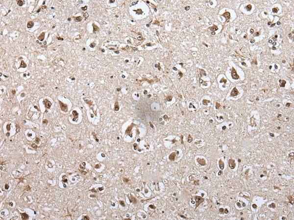
Immunohistochemical staining of human cerebellum shows strong nuclear positivity in Purkinje cells.
 and METAP1D over-expression lysate (Co-expressed with a C-terminal myc-DDK tag (~3.1 kDa) in mammalian HEK293T cells, LY404696).")
Immunohistochemical staining of human cerebellum shows strong nuclear positivity in Purkinje cells.
Anti-METAP1D Antibody
HPA030298
ApplicationsWestern Blot, ImmunoCytoChemistry, ImmunoHistoChemistry
Product group Antibodies
ReactivityHuman
TargetMETAP1D
Overview
- SupplierAtlas Antibodies
- Product NameAnti-METAP1D Antibody
- Delivery Days Customer4
- ApplicationsWestern Blot, ImmunoCytoChemistry, ImmunoHistoChemistry
- CertificationResearch Use Only
- ClonalityPolyclonal
- ConjugateUnconjugated
- Gene ID254042
- Target nameMETAP1D
- Target descriptionmethionyl aminopeptidase type 1D, mitochondrial
- Target synonymsMAP 1D, MAP1D, MetAP 1D, Metap1l, methionine aminopeptidase 1D, mitochondrial, CDS of metAP-3 within PCR fragment, metAP 1D, peptidase M 1D
- HostRabbit
- IsotypeIgG
- Protein IDQ6UB28
- Protein NameMethionine aminopeptidase 1D, mitochondrial
- Scientific DescriptionRecombinant Protein Epitope Signature Tag (PrEST) antigen sequence
- ReactivityHuman
- Storage Instruction-20°C,2°C to 8°C
- UNSPSC41116161
Datasheet
MSDS
Related products
Product group Antibodies
Anti-METAP1D AntibodyA45268
ApplicationsImmunoHistoChemistry
ReactivityHuman
- SizePrice

Product group Antibodies
Anti-METAP1D Antibody Picoband(r)A11898-1-CARRIER-FREE
ApplicationsWestern Blot, ELISA, ImmunoHistoChemistry
ReactivityHuman, Mouse, Rat
TargetMETAP1D
- SizePrice
Product group Antibodies
Anti-MAP1D (N-term) Antibody102-20651
ApplicationsWestern Blot
TargetMETAP1D
- SizePrice
Product group Antibodies

MAP1D AntibodyLS-C830338
ApplicationsELISA, ImmunoHistoChemistry
ReactivityHuman, Mouse
TargetMETAP1D
- SizePrice
Product group Antibodies
MAP1D AntibodyCSB-PA861884XA01DOA
ApplicationsWestern Blot, ELISA
ReactivityPlant
TargetMAP1D
- SizePrice

Product group Antibodies
Anti-METAP1D AntibodyHPA030299
ApplicationsWestern Blot, ImmunoCytoChemistry, ImmunoHistoChemistry
ReactivityHuman
TargetMETAP1D
- SizePrice