


Immunohistochemical staining of human cerebral cortex shows strong cytoplasmic positivity in neuronal cells.

Immunohistochemical staining of human cerebral cortex shows strong cytoplasmic positivity in neuronal cells.
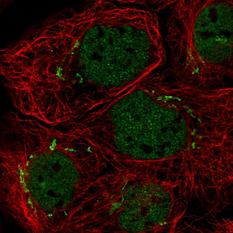
Anti-MFAP3 Antibody
HPA015883
ApplicationsWestern Blot, ImmunoHistoChemistry
Product group Antibodies
ReactivityHuman
TargetMFAP3
Overview
- SupplierAtlas Antibodies
- Product NameAnti-MFAP3 Antibody
- Delivery Days Customer4
- ApplicationsWestern Blot, ImmunoHistoChemistry
- CertificationResearch Use Only
- ClonalityPolyclonal
- ConjugateUnconjugated
- Gene ID4238
- Target nameMFAP3
- Target descriptionmicrofibril associated protein 3
- Target synonymsmicrofibril-associated glycoprotein 3, microfibrillar associated protein 3
- HostRabbit
- IsotypeIgG
- Protein IDP55082
- Protein NameMicrofibril-associated glycoprotein 3
- Scientific DescriptionRecombinant Protein Epitope Signature Tag (PrEST) antigen sequence
- ReactivityHuman
- Storage Instruction-20°C,2°C to 8°C
- UNSPSC41116161
Datasheet
MSDS
Related products
Product group Antibodies
ApplicationsImmunoPrecipitation, Western Blot, ImmunoCytoChemistry, ImmunoHistoChemistry
TargetMFAP3
- SizePrice
Product group Antibodies

MFAP3 Antibody (Biotin)LS-C395938
ApplicationsELISA
ReactivityHuman
TargetMFAP3
- SizePrice
Product group Antibodies
Anti-MFAP3 AntibodyHPA049198
ApplicationsImmunoCytoChemistry
ReactivityHuman
TargetMFAP3
- SizePrice
Product group Antibodies
Anti-MFAP3 AntibodyHPA049198
ApplicationsImmunoCytoChemistry
ReactivityHuman
TargetMFAP3
- SizePrice
Product group Antibodies
MFAP3 AntibodyCSB-PA013747LA01HU
ApplicationsELISA
ReactivityHuman
TargetMFAP3
- SizePrice

Product group Antibodies
Anti-MFAP3 Antibody Picoband(r)A13942-CARRIER-FREE
ApplicationsFlow Cytometry, Western Blot, ELISA, ImmunoHistoChemistry
ReactivityHuman, Mouse, Rat
TargetMFAP3
- SizePrice