Anti Microtubule-Associated Proteins 1A/1B Light Chain 3A (MAP1LC3A/LC3) mAb (Clone LC3-1703)
CAC-CTB-LC3-2-IC
Product group Antibodies
Overview
- SupplierCosmo Bio USA
- Product NameAnti Microtubule-Associated Proteins 1A/1B Light Chain 3A (MAP1LC3A/LC3) mAb (Clone LC3-1703)
- Delivery Days Customer16
- CertificationResearch Use Only
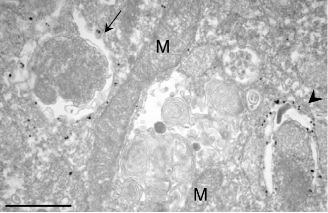
- Scientific DescriptionLC3 is a mammalian autophagosome marker. It is immediately cleaved to form LC3-I by Atg4, and when autophagy is induced, phosphatidylethanolamine is covalently linked to LC3-I C-terminal glycine to form LC3-II. LC3-II is membrane-bound and thought to be present in autophagosomal membranes. Since autophagosomes are degraded by fusion with lysosomes, LC3-II itself is also degraded by autophagy. Therefore, it is generally accepted that the amount of LC3-II correlates well with the amount of autophagosomes. This antibody shows good results in ICC and Immunoelectron microscopy experiments. Source: Professor Noboru Mizushima, Department of Cell Physiology, Tokyo Medical and Dental University References: 1) Kabeya, Y., Mizushima, N., Ueno, T., Yamamoto, A., Kirisako, T., Noda, T., Kominami, E., Ohsumi, Y. and Yoshimori, T. LC3, a mammalian homologue of yeast Apg8p, is localized in autophagosome membranes after processing EMBO J. 19, 5720-5728. (2000) 2) Mizushima, N., Yoshimori, T. How to interpret LC3 immunoblotting Autophagy 3:542-545 (2007) 3) Mizushima, N., Yoshimori, T. and Levine, B. Methods in mammalian autophagy research. Cell 140; 313-326 (2010)
- UNSPSC12352203

