


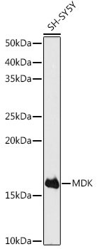
Figure 1. Western blot analysis of MDK using anti-MDK antibody (RP1051). Electrophoresis was performed on a 5-20% SDS-PAGE gel at 70V (Stacking gel) / 90V (Resolving gel) for 2-3 hours. The sample well of each lane was loaded with 30 ug of sample under reducing conditions. Lane 1: human SH-SY5Y whole cell lysates, Lane 2: human U20S whole cell lysates. After electrophoresis, proteins were transferred to a nitrocellulose membrane at 150 mA for 50-90 minutes. Blocked the membrane with 5% non-fat milk/TBS for 1.5 hour at RT. The membrane was incubated with rabbit anti-MDK antigen affinity purified polyclonal antibody (Catalog # RP1051) at 0.5 microg/mL overnight at 4°C, then washed with TBS-0.1%Tween 3 times with 5 minutes each and probed with a goat anti-rabbit IgG-HRP secondary antibody at a dilution of 1:5000 for 1.5 hour at RT. The signal is developed using an Enhanced Chemiluminescent detection (ECL) kit (Catalog # EK1002) with Tanon 5200 system. A specific band was detected for MDK at approximately 16 kDa. The expected band size for MDK is at 16 kDa.
Figure 1. Western blot analysis of MDK using anti-MDK antibody (RP1051). Electrophoresis was performed on a 5-20% SDS-PAGE gel at 70V (Stacking gel) / 90V (Resolving gel) for 2-3 hours. The sample well of each lane was loaded with 30 ug of sample under reducing conditions. Lane 1: human SH-SY5Y whole cell lysates, Lane 2: human U20S whole cell lysates. After electrophoresis, proteins were transferred to a nitrocellulose membrane at 150 mA for 50-90 minutes. Blocked the membrane with 5% non-fat milk/TBS for 1.5 hour at RT. The membrane was incubated with rabbit anti-MDK antigen affinity purified polyclonal antibody (Catalog # RP1051) at 0.5 microg/mL overnight at 4°C, then washed with TBS-0.1%Tween 3 times with 5 minutes each and probed with a goat anti-rabbit IgG-HRP secondary antibody at a dilution of 1:5000 for 1.5 hour at RT. The signal is developed using an Enhanced Chemiluminescent detection (ECL) kit (Catalog # EK1002) with Tanon 5200 system. A specific band was detected for MDK at approximately 16 kDa. The expected band size for MDK is at 16 kDa.
Anti-Midkine/MDK Antibody Picoband(r)
RP1051-CARRIER-FREE
ApplicationsWestern Blot
Product group Antibodies
ReactivityHuman
TargetMDK
Overview
- SupplierBoster Bio
- Product NameAnti-Midkine/MDK Antibody Picoband(r)
- Delivery Days Customer9
- Application Supplier NoteWB: The detection limit for Midkine is approximately 0.25ng/lane under reducing conditions. Tested Species: In-house tested species with positive results. Other applications have not been tested. Optimal dilutions should be determined by end users.
- ApplicationsWestern Blot
- CertificationResearch Use Only
- ClonalityPolyclonal
- Concentration500 ug/ml
- Gene ID4192
- Target nameMDK
- Target descriptionmidkine
- Target synonymsARAP, MK, NEGF2, midkine, amphiregulin-associated protein, midgestation and kidney protein, neurite growth-promoting factor 2, neurite outgrowth-promoting factor 2, retinoic acid inducible factor
- HostRabbit
- IsotypeIgG
- Protein IDP21741
- Protein NameMidkine
- Scientific DescriptionBoster Bio Anti-Midkine/MDK Antibody catalog # RP1051. Tested in WB applications. This antibody reacts with Human. The brand Picoband indicates this is a premium antibody that guarantees superior quality, high affinity, and strong signals with minimal background in Western blot applications. Only our best-performing antibodies are designated as Picoband, ensuring unmatched performance.
- ReactivityHuman
- Storage Instruction-20°C,2°C to 8°C
- UNSPSC12352203
Related products
Product group Antibodies
Anti-MDK (C-term) Antibody102-21018
ApplicationsImmunoFluorescence, Western Blot, ImmunoHistoChemistry, ImmunoHistoChemistry Paraffin
TargetMDK
- SizePrice
Product group Antibodies
Anti-Midkine AntibodyA12574
ApplicationsImmunoFluorescence, Western Blot, ImmunoCytoChemistry
ReactivityHuman, Mouse, Rat
- SizePrice
Product group Antibodies
Midkine Recombinant AntibodyBSM-61219R
ApplicationsImmunoFluorescence, ImmunoPrecipitation, Western Blot, ImmunoCytoChemistry, ImmunoHistoChemistry, ImmunoHistoChemistry Frozen, ImmunoHistoChemistry Paraffin
TargetMDK
- SizePrice
Product group Antibodies
MDK AntibodyCSB-PA003244
ApplicationsWestern Blot, ELISA
ReactivityHuman, Rat
TargetMDK
- SizePrice
Product group Antibodies
Mdk Polyclonal AntibodyCAC09128
ApplicationsImmunoFluorescence, ELISA, ImmunoHistoChemistry
TargetMDK
- SizePrice
Product group Antibodies

ApplicationsWestern Blot, ELISA
ReactivityHuman
TargetMDK
- SizePrice
Product group Antibodies
Midkine antibodyGTX12739
ApplicationsWestern Blot, ELISA
ReactivityHuman
- SizePrice
Product group Antibodies
Anti-MDK AntibodyCAB0251
ApplicationsImmunoFluorescence, Western Blot, ELISA, ImmunoCytoChemistry
ReactivityHuman
TargetMDK
- SizePrice