


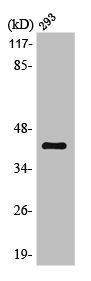
Figure 1. Western blot analysis of MKK6/MAP2K6 using anti-MKK6/MAP2K6 antibody (A02011-3). Electrophoresis was performed on a 5-20% SDS-PAGE gel at 70V (Stacking gel) / 90V (Resolving gel) for 2-3 hours. The sample well of each lane was loaded with 30 ug of sample under reducing conditions. Lane 1: rat kidney tissue lysates, Lane 2: rat skeletal muscle tissue lysates, Lane 3: mouse skeletal muscle tissue lysates. After electrophoresis, proteins were transferred to a nitrocellulose membrane at 150 mA for 50-90 minutes. Blocked the membrane with 5% non-fat milk/TBS for 1.5 hour at RT. The membrane was incubated with rabbit anti-MKK6/MAP2K6 antigen affinity purified polyclonal antibody (Catalog # A02011-3) at 0.5 microg/mL overnight at 4°C, then washed with TBS-0.1%Tween 3 times with 5 minutes each and probed with a goat anti-rabbit IgG-HRP secondary antibody at a dilution of 1:5000 for 1.5 hour at RT. The signal is developed using an Enhanced Chemiluminescent detection (ECL) kit (Catalog # EK1002) with Tanon 5200 system. A specific band was detected for MKK6/MAP2K6 at approximately 38 kDa. The expected band size for MKK6/MAP2K6 is at 38 kDa.
Figure 1. Western blot analysis of MKK6/MAP2K6 using anti-MKK6/MAP2K6 antibody (A02011-3). Electrophoresis was performed on a 5-20% SDS-PAGE gel at 70V (Stacking gel) / 90V (Resolving gel) for 2-3 hours. The sample well of each lane was loaded with 30 ug of sample under reducing conditions. Lane 1: rat kidney tissue lysates, Lane 2: rat skeletal muscle tissue lysates, Lane 3: mouse skeletal muscle tissue lysates. After electrophoresis, proteins were transferred to a nitrocellulose membrane at 150 mA for 50-90 minutes. Blocked the membrane with 5% non-fat milk/TBS for 1.5 hour at RT. The membrane was incubated with rabbit anti-MKK6/MAP2K6 antigen affinity purified polyclonal antibody (Catalog # A02011-3) at 0.5 microg/mL overnight at 4°C, then washed with TBS-0.1%Tween 3 times with 5 minutes each and probed with a goat anti-rabbit IgG-HRP secondary antibody at a dilution of 1:5000 for 1.5 hour at RT. The signal is developed using an Enhanced Chemiluminescent detection (ECL) kit (Catalog # EK1002) with Tanon 5200 system. A specific band was detected for MKK6/MAP2K6 at approximately 38 kDa. The expected band size for MKK6/MAP2K6 is at 38 kDa.
Anti-MKK6/MAP2K6 Antibody Picoband(r)
A02011-3-DYLIGHT594
ApplicationsWestern Blot, ELISA
Product group Antibodies
ReactivityHuman, Mouse, Rat
TargetMAP2K6
Overview
- SupplierBoster Bio
- Product NameAnti-MKK6/MAP2K6 Antibody Picoband(r)
- Delivery Days Customer9
- Application Supplier NoteTested Species: In-house tested species with positive results. Predicted Species: Species predicted to be fit for the product based on sequence similarities. Other applications have not been tested. Optimal dilutions should be determined by end users.
- ApplicationsWestern Blot, ELISA
- CertificationResearch Use Only
- ClonalityPolyclonal
- Concentration500 ug/ml
- ConjugateOther Conjugate
- Gene ID5608
- Target nameMAP2K6
- Target descriptionmitogen-activated protein kinase kinase 6
- Target synonymsCRCMSL, MAPKK6, MEK6, MKK6, PRKMK6, SAPKK-3, SAPKK3, dual specificity mitogen-activated protein kinase kinase 6, Colorectal Cancer Metastasis-Suppressed LncRNA, MAPK/ERK kinase 6, MAPKK 6, MEK 6, SAPK kinase 3, protein kinase, mitogen-activated, kinase 6 (MAP kinase kinase 6), stress-activated protein kinase kinase 3
- HostRabbit
- IsotypeIgG
- Protein IDP52564
- Protein NameDual specificity mitogen-activated protein kinase kinase 6
- Scientific DescriptionBoster Bio Anti-MKK6/MAP2K6 Antibody Picoband® catalog # A02011-3. Tested in ELISA, WB applications. This antibody reacts with Human, Mouse, Rat. The brand Picoband indicates this is a premium antibody that guarantees superior quality, high affinity, and strong signals with minimal background in Western blot applications. Only our best-performing antibodies are designated as Picoband, ensuring unmatched performance.
- ReactivityHuman, Mouse, Rat
- Storage Instruction-20°C,2°C to 8°C
- UNSPSC12352203
Related products
Product group Antibodies
Anti-MAP2K6 Antibody144-65549
ApplicationsImmunoFluorescence, Western Blot
ReactivityHuman, Mouse, Rat
TargetMAP2K6
- SizePrice
Product group Antibodies
ApplicationsWestern Blot, ELISA, ImmunoCytoChemistry, ImmunoHistoChemistry, ImmunoHistoChemistry Frozen, ImmunoHistoChemistry Paraffin
ReactivityMouse
TargetMAP2K6
- SizePrice
Product group Antibodies
MEK6 Polyclonal AntibodyBS-2911R
ApplicationsImmunoFluorescence, Western Blot, ELISA, ImmunoCytoChemistry, ImmunoHistoChemistry, ImmunoHistoChemistry Frozen, ImmunoHistoChemistry Paraffin
ReactivityBovine, Canine, Human, Mouse, Porcine, Rabbit, Rat
TargetMAP2K6
- SizePrice
Product group Antibodies
Anti-MKK6 AntibodyA96155
ApplicationsWestern Blot, ELISA, ImmunoHistoChemistry
ReactivityHuman, Mouse, Rat
- SizePrice
Product group Antibodies

MAP2K6 / MEK6 / MKK6 AntibodyLS-C831451
ApplicationsImmunoHistoChemistry
ReactivityHuman, Mouse, Rat
TargetMAP2K6
- SizePrice
Product group Antibodies
MAP2K6 AntibodyCSB-PA003224
ApplicationsImmunoFluorescence, Western Blot, ELISA, ImmunoHistoChemistry
ReactivityHuman, Mouse, Rat
TargetMAP2K6
- SizePrice

Product group Antibodies
Anti-MAP2K6 AntibodyHPA031134
ApplicationsWestern Blot, ImmunoCytoChemistry, ImmunoHistoChemistry
ReactivityHuman, Mouse, Rat
TargetMAP2K6
- SizePrice
![Whole cell extract (30 μg) was separated by 10% SDS-PAGE, and the membrane was blotted with MEK6 antibody [N1C3] (GTX107445) diluted at 1:1000. The HRP-conjugated anti-rabbit IgG antibody (GTX213110-01) was used to detect the primary antibody.](https://www.genetex.com/upload/website/prouct_img/normal/GTX107445/GTX107445_40037_20220401_WB_2_w_23060120_679.webp)
Product group Antibodies
References
MEK6 antibody [N1C3]GTX107445
ApplicationsImmunoFluorescence, Western Blot, ImmunoCytoChemistry, ImmunoHistoChemistry, ImmunoHistoChemistry Paraffin
ReactivityHuman, Mouse
TargetMAP2K6
- SizePrice