


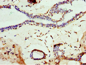
Immunohistochemical staining of rat cerebellum shows strong immunoreactivity in myelinated neural fibers.
![Lane 1: Marker [kDa] 250, 130, 100, 70, 55, 35, 25, 15, 10. Lane 2: Human Cerebral Cortex tissue](https://atlasantibodies.s3.amazonaws.com/images/wb/amab91066-wb-1.jpg "Lane 1: Marker [kDa] 250, 130, 100, 70, 55, 35, 25, 15, 10. Lane 2: Human Cerebral Cortex tissue")
Immunohistochemical staining of rat cerebellum shows strong immunoreactivity in myelinated neural fibers.
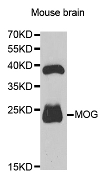
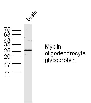
Anti-MOG Antibody
AMAB91066
ApplicationsWestern Blot, ImmunoHistoChemistry
Product group Antibodies
ReactivityHuman, Mouse, Rat
TargetMOG
Overview
- SupplierAtlas Antibodies
- Product NameAnti-MOG Antibody
- Delivery Days Customer4
- ApplicationsWestern Blot, ImmunoHistoChemistry
- CertificationResearch Use Only
- ClonalityMonoclonal
- Clone IDCL2852
- ConjugateUnconjugated
- Gene ID4340
- Target nameMOG
- Target descriptionmyelin oligodendrocyte glycoprotein
- Target synonymsBTN6, BTNL11, MOGIG2, NRCLP7, myelin-oligodendrocyte glycoprotein, MOG AluA, MOG AluB, MOG Ig-AluB, MOG alpha-5
- HostMouse
- IsotypeIgG1
- Protein IDQ16653
- Protein NameMyelin-oligodendrocyte glycoprotein
- Scientific DescriptionRecombinant Protein Epitope Signature Tag (PrEST) antigen sequence
- ReactivityHuman, Mouse, Rat
- Storage Instruction-20°C,2°C to 8°C
- UNSPSC41116161
Datasheet
MSDS
Related products
Product group Antibodies
Anti-MOG AntibodyA35668
ApplicationsImmunoFluorescence, Western Blot, ImmunoHistoChemistry
ReactivityHuman, Mouse, Rat
- SizePrice

Product group Antibodies
Anti-Myelin oligodendrocyte glycoprotein/MOG Antibody Picoband(r)A03294-CARRIER-FREE
ApplicationsFlow Cytometry, Western Blot, ImmunoHistoChemistry
ReactivityHuman, Mouse, Rat
TargetMOG
- SizePrice
Product group Antibodies
Anti-MOG IgG Antibody130-10907-20
ApplicationsELISA
ReactivityHuman
TargetMOG
- SizePrice

Product group Antibodies
Anti-MOG AntibodyAMAB91067
ApplicationsWestern Blot, ImmunoHistoChemistry
ReactivityHuman, Mouse, Rat
TargetMOG
- SizePrice
Product group Antibodies

MOG AntibodyLS-C834971
ApplicationsWestern Blot, ELISA, ImmunoHistoChemistry
ReactivityHuman, Mouse, Rat
TargetMOG
- SizePrice
Product group Antibodies
MOG Polyclonal AntibodyBS-0426R
ApplicationsFlow Cytometry, ImmunoFluorescence, Western Blot, ELISA, ImmunoCytoChemistry, ImmunoHistoChemistry, ImmunoHistoChemistry Frozen, ImmunoHistoChemistry Paraffin
ReactivityGuinea Pig, Human, Mouse, Porcine, Rat
TargetMOG
- SizePrice
Product group Antibodies

Goat anti-MOGEB06668
ApplicationsWestern Blot, ELISA
ReactivityBovine, Human, Mouse, Porcine, Rat
TargetMOG
- SizePrice
Product group Antibodies
MOG AntibodyCSB-PA619083ESR2HU
ApplicationsELISA, ImmunoHistoChemistry
ReactivityHuman
TargetMOG
- SizePrice
Product group Antibodies
Mog Polyclonal AntibodyCAC10670
ApplicationsELISA, ImmunoHistoChemistry
TargetMOG
- SizePrice