

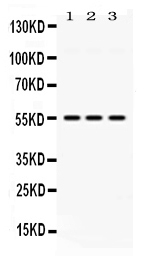
Figure 1. Western blot analysis of MPP1 using anti-MPP1 antibody (PB10078). Electrophoresis was performed on a 5-20% SDS-PAGE gel at 70V (Stacking gel) / 90V (Resolving gel) for 2-3 hours. The sample well of each lane was loaded with 30 ug of sample under reducing conditions. Lane 1: rat lung tissue lysates, Lane 2: mouse spleen tissue lysates, Lane 3: MCF-7 whole cell lysates. After electrophoresis, proteins were transferred to a nitrocellulose membrane at 150 mA for 50-90 minutes. Blocked the membrane with 5% non-fat milk/TBS for 1.5 hour at RT. The membrane was incubated with rabbit anti-MPP1 antigen affinity purified polyclonal antibody (Catalog # PB10078) at 0.5 microg/mL overnight at 4°C, then washed with TBS-0.1%Tween 3 times with 5 minutes each and probed with a goat anti-rabbit IgG-HRP secondary antibody at a dilution of 1:5000 for 1.5 hour at RT. The signal is developed using an Enhanced Chemiluminescent detection (ECL) kit (Catalog # EK1002) with Tanon 5200 system. A specific band was detected for MPP1 at approximately 55 kDa. The expected band size for MPP1 is at 52 kDa.
. MPP1 was detected in immunocytochemical section of MCF7 cells. Enzyme antigen retrieval was performed using IHC enzyme antigen retrieval reagent (AR0022) for 15 mins. The cells were blocked with 10% goat serum. And then incubated with 2microg/mL rabbit anti-MPP1 Antibody (PB10078) overnight at 4°C. DyLight®594 Conjugated Goat Anti-Rabbit IgG (BA1142) was used as secondary antibody at 1:100 dilution and incubated for 30 minutes at 37°C. The section was counterstained with DAPI. Visualize using a fluorescence microscope and filter sets appropriate for the label used.")
Figure 1. Western blot analysis of MPP1 using anti-MPP1 antibody (PB10078). Electrophoresis was performed on a 5-20% SDS-PAGE gel at 70V (Stacking gel) / 90V (Resolving gel) for 2-3 hours. The sample well of each lane was loaded with 30 ug of sample under reducing conditions. Lane 1: rat lung tissue lysates, Lane 2: mouse spleen tissue lysates, Lane 3: MCF-7 whole cell lysates. After electrophoresis, proteins were transferred to a nitrocellulose membrane at 150 mA for 50-90 minutes. Blocked the membrane with 5% non-fat milk/TBS for 1.5 hour at RT. The membrane was incubated with rabbit anti-MPP1 antigen affinity purified polyclonal antibody (Catalog # PB10078) at 0.5 microg/mL overnight at 4°C, then washed with TBS-0.1%Tween 3 times with 5 minutes each and probed with a goat anti-rabbit IgG-HRP secondary antibody at a dilution of 1:5000 for 1.5 hour at RT. The signal is developed using an Enhanced Chemiluminescent detection (ECL) kit (Catalog # EK1002) with Tanon 5200 system. A specific band was detected for MPP1 at approximately 55 kDa. The expected band size for MPP1 is at 52 kDa.
Anti-MPP1 Antibody Picoband(r)
PB10078-CARRIER-FREE
ApplicationsImmunoFluorescence, Western Blot, ImmunoCytoChemistry
Product group Antibodies
ReactivityHamster, Human, Mouse, Rat
TargetMPP1
Overview
- SupplierBoster Bio
- Product NameAnti-MPP1 Antibody Picoband(r)
- Delivery Days Customer9
- Application Supplier NoteTested Species: In-house tested species with positive results. Other applications have not been tested. Optimal dilutions should be determined by end users.
- ApplicationsImmunoFluorescence, Western Blot, ImmunoCytoChemistry
- CertificationResearch Use Only
- ClonalityPolyclonal
- Concentration500 ug/ml
- Gene ID4354
- Target nameMPP1
- Target descriptionMAGUK p55 scaffold protein 1
- Target synonymsAAG12, DXS552E, EMP55, MRG1, PEMP, 55 kDa erythrocyte membrane protein, aging-associated gene 12, erythrocyte membrane protein p55, membrane palmitoylated protein 1, membrane protein, palmitoylated 1, 55kDa, migration-related gene 1, p55, palmitoylated erythrocyte membrane protein
- HostRabbit
- IsotypeIgG
- Protein IDQ00013
- Protein Name55 kDa erythrocyte membrane protein
- Scientific DescriptionBoster Bio Anti-MPP1 Antibody Picoband® catalog # PB10078. Tested in IF, ICC, WB applications. This antibody reacts with Human, Mouse, Rat. The brand Picoband indicates this is a premium antibody that guarantees superior quality, high affinity, and strong signals with minimal background in Western blot applications. Only our best-performing antibodies are designated as Picoband, ensuring unmatched performance.
- ReactivityHamster, Human, Mouse, Rat
- Storage Instruction-20°C,2°C to 8°C
- UNSPSC12352203
Related products
Product group Antibodies
Anti-MPP1 AntibodyA31276
ApplicationsWestern Blot, ImmunoHistoChemistry
ReactivityHuman, Mouse
- SizePrice
Product group Antibodies
Anti-MPP1 Antibody144-06298
ApplicationsWestern Blot
ReactivityHuman, Mouse
TargetMPP1
- SizePrice
Product group Antibodies

MPP1 AntibodyLS-C830808
ApplicationsELISA, ImmunoHistoChemistry
ReactivityHuman, Mouse
TargetMPP1
- SizePrice
Product group Antibodies
MPP1 Polyclonal AntibodyBS-9522R
ApplicationsWestern Blot
ReactivityBovine, Canine, Human, Mouse, Porcine, Rat, Sheep
TargetMPP1
- SizePrice
Product group Antibodies
MPP1 AntibodyCSB-PA014758LA01HU
ApplicationsELISA, ImmunoHistoChemistry
ReactivityHuman
TargetMPP1
- SizePrice

Product group Antibodies
Anti-MPP1 AntibodyHPA000167
ApplicationsImmunoCytoChemistry
ReactivityHuman
TargetMPP1
- SizePrice
Product group Antibodies
MPP1 antibody [N3C3]GTX111341
ApplicationsWestern Blot, ImmunoHistoChemistry, ImmunoHistoChemistry Paraffin
ReactivityHuman
TargetMPP1
- SizePrice
Product group Antibodies
Anti-MPP1 AntibodyCAB6298
ApplicationsWestern Blot, ELISA
ReactivityHuman
TargetMPP1
- SizePrice