


Immunohistochemical staining of human cerebral cortex shows moderate cytoplasmic positivity in neuronal cells.
 and MPP2 over-expression lysate (Co-expressed with a C-terminal myc-DDK tag (~3.1 kDa) in mammalian HEK293T cells, LY417352).")
Immunohistochemical staining of human cerebral cortex shows moderate cytoplasmic positivity in neuronal cells.
Anti-MPP2 Antibody
HPA026486
ApplicationsWestern Blot, ImmunoCytoChemistry, ImmunoHistoChemistry
Product group Antibodies
ReactivityHuman
TargetMPP2
Overview
- SupplierAtlas Antibodies
- Product NameAnti-MPP2 Antibody
- Delivery Days Customer4
- ApplicationsWestern Blot, ImmunoCytoChemistry, ImmunoHistoChemistry
- CertificationResearch Use Only
- ClonalityPolyclonal
- ConjugateUnconjugated
- Gene ID4355
- Target nameMPP2
- Target descriptionMAGUK p55 scaffold protein 2
- Target synonymsDLG2, MAGUK p55 subfamily member 2, discs large, homolog 2, membrane palmitoylated protein 2, membrane protein, palmitoylated 2 (MAGUK p55 subfamily member 2)
- HostRabbit
- IsotypeIgG
- Protein IDQ14168
- Protein NameMAGUK p55 subfamily member 2
- Scientific DescriptionRecombinant Protein Epitope Signature Tag (PrEST) antigen sequence
- ReactivityHuman
- Storage Instruction-20°C,2°C to 8°C
- UNSPSC41116161
Datasheet
MSDS
Related products
Product group Antibodies
Anti-MPP2 AntibodyA31498
ApplicationsWestern Blot, ImmunoHistoChemistry
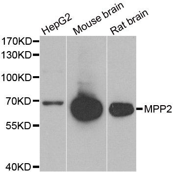
ReactivityHuman, Mouse, Rat
- SizePrice
Product group Antibodies
MPP2 Polyclonal AntibodyBS-13689R
ApplicationsImmunoFluorescence, ELISA, ImmunoCytoChemistry, ImmunoHistoChemistry, ImmunoHistoChemistry Frozen, ImmunoHistoChemistry Paraffin
ReactivityBovine, Canine, Equine, Human, Mouse, Rabbit, Rat
- SizePrice
Product group Antibodies
ApplicationsImmunoPrecipitation, Western Blot, ImmunoCytoChemistry, ImmunoHistoChemistry
ReactivityMouse, Porcine, Rat
TargetMPP2
- SizePrice
Product group Antibodies
MPP2 antibody [N1C3]GTX103908
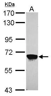
ApplicationsWestern Blot
ReactivityHuman, Mouse
TargetMPP2
- SizePrice
Product group Antibodies

MPP2 AntibodyLS-C334856
ApplicationsImmunoFluorescence, Western Blot, ImmunoHistoChemistry
ReactivityHuman, Mouse, Rat
TargetMPP2
- SizePrice

Product group Antibodies
Anti-MPP2 AntibodyHPA073483
ApplicationsImmunoHistoChemistry
ReactivityHuman
TargetMPP2
- SizePrice

Product group Antibodies
Anti-MPP2 Antibody Picoband(r)PB10079-CARRIER-FREE
ApplicationsWestern Blot
ReactivityHuman, Mouse, Rat
TargetMPP2
- SizePrice