


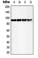
Figure 1. Western blot analysis of MSK1 using anti-MSK1 antibody (PB9268). Electrophoresis was performed on a 5-20% SDS-PAGE gel at 70V (Stacking gel) / 90V (Resolving gel) for 2-3 hours. The sample well of each lane was loaded with 30 ug of sample under reducing conditions. Lane 1: human HEL whole cell lysates, Lane 2: rat brain tissue lysates, Lane 3: mouse brain tissue lysates. After electrophoresis, proteins were transferred to a nitrocellulose membrane at 150 mA for 50-90 minutes. Blocked the membrane with 5% non-fat milk/TBS for 1.5 hour at RT. The membrane was incubated with rabbit anti-MSK1 antigen affinity purified polyclonal antibody (Catalog # PB9268) at 0.5 microg/mL overnight at 4°C, then washed with TBS-0.1%Tween 3 times with 5 minutes each and probed with a goat anti-rabbit IgG-HRP secondary antibody at a dilution of 1:5000 for 1.5 hour at RT. The signal is developed using an Enhanced Chemiluminescent detection (ECL) kit (Catalog # EK1002) with Tanon 5200 system. A specific band was detected for MSK1 at approximately 90 kDa. The expected band size for MSK1 is at 90 kDa.
. MSK1 was detected in a paraffin-embedded section of Mouse Cardiac Muscle tissue. Heat mediated antigen retrieval was performed in EDTA buffer (pH 8.0, epitope retrieval solution). The tissue section was blocked with 10% goat serum. The tissue section was then incubated with 1 microg/ml rabbit anti-MSK1 Antibody (PB9268) overnight at 4°C. Biotinylated goat anti-rabbit IgG was used as secondary antibody and incubated for 30 minutes at 37°C. The tissue section was developed using Strepavidin-Biotin-Complex (SABC) (Catalog # SA1022) with DAB as the chromogen.")
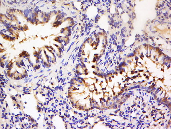
. MSK1 was detected in a paraffin-embedded section of Rat Brain tissue. Heat mediated antigen retrieval was performed in EDTA buffer (pH 8.0, epitope retrieval solution). The tissue section was blocked with 10% goat serum. The tissue section was then incubated with 1 microg/ml rabbit anti-MSK1 Antibody (PB9268) overnight at 4°C. Biotinylated goat anti-rabbit IgG was used as secondary antibody and incubated for 30 minutes at 37°C. The tissue section was developed using Strepavidin-Biotin-Complex (SABC) (Catalog # SA1022) with DAB as the chromogen.")
. MSK1 was detected in a paraffin-embedded section of Human Placenta tissue. Heat mediated antigen retrieval was performed in EDTA buffer (pH 8.0, epitope retrieval solution). The tissue section was blocked with 10% goat serum. The tissue section was then incubated with 1 microg/ml rabbit anti-MSK1 Antibody (PB9268) overnight at 4°C. Biotinylated goat anti-rabbit IgG was used as secondary antibody and incubated for 30 minutes at 37°C. The tissue section was developed using Strepavidin-Biotin-Complex (SABC) (Catalog # SA1022) with DAB as the chromogen.")
. MSK1 was detected in an immunocytochemical section of T47D cells. Enzyme antigen retrieval was performed using IHC enzyme antigen retrieval reagent (AR0022) for 15 mins. The cells were blocked with 10% goat serum. And then incubated with 5 microg/mL rabbit anti-MSK1 Antibody (PB9268) overnight at 4°C. DyLight®488 Conjugated Goat Anti-Rabbit IgG (BA1127) was used as secondary antibody at 1:500 dilution and incubated for 30 minutes at 37°C. The section was counterstained with DAPI. Visualize using a fluorescence microscope and filter sets appropriate for the label used.")
Figure 1. Western blot analysis of MSK1 using anti-MSK1 antibody (PB9268). Electrophoresis was performed on a 5-20% SDS-PAGE gel at 70V (Stacking gel) / 90V (Resolving gel) for 2-3 hours. The sample well of each lane was loaded with 30 ug of sample under reducing conditions. Lane 1: human HEL whole cell lysates, Lane 2: rat brain tissue lysates, Lane 3: mouse brain tissue lysates. After electrophoresis, proteins were transferred to a nitrocellulose membrane at 150 mA for 50-90 minutes. Blocked the membrane with 5% non-fat milk/TBS for 1.5 hour at RT. The membrane was incubated with rabbit anti-MSK1 antigen affinity purified polyclonal antibody (Catalog # PB9268) at 0.5 microg/mL overnight at 4°C, then washed with TBS-0.1%Tween 3 times with 5 minutes each and probed with a goat anti-rabbit IgG-HRP secondary antibody at a dilution of 1:5000 for 1.5 hour at RT. The signal is developed using an Enhanced Chemiluminescent detection (ECL) kit (Catalog # EK1002) with Tanon 5200 system. A specific band was detected for MSK1 at approximately 90 kDa. The expected band size for MSK1 is at 90 kDa.
Anti-MSK1/RPS6KA5 Antibody Picoband(r)
PB9268-CARRIER-FREE
ApplicationsImmunoFluorescence, Western Blot, ImmunoCytoChemistry, ImmunoHistoChemistry
Product group Antibodies
ReactivityHuman, Mouse, Rat
TargetRPS6KA5
Overview
- SupplierBoster Bio
- Product NameAnti-MSK1/RPS6KA5 Antibody Picoband(r)
- Delivery Days Customer9
- Application Supplier NoteWB: The detection limit for MSK1 is approximately 0.25ng/lane under reducing conditions. Tested Species: In-house tested species with positive results. By Heat: Boiling the paraffin sections in 10mM citrate buffer, pH6.0, for 20mins is required for the staining of formalin/paraffin sections. Other applications have not been tested. Optimal dilutions should be determined by end users.
- ApplicationsImmunoFluorescence, Western Blot, ImmunoCytoChemistry, ImmunoHistoChemistry
- CertificationResearch Use Only
- ClonalityPolyclonal
- Concentration500 ug/ml
- Gene ID9252
- Target nameRPS6KA5
- Target descriptionribosomal protein S6 kinase A5
- Target synonymsMSK1, MSPK1, RLPK, ribosomal protein S6 kinase alpha-5, 90 kDa ribosomal protein S6 kinase 5, RSK-like protein kinase, RSKL, S6K-alpha-5, nuclear mitogen- and stress-activated protein kinase 1, ribosomal protein S6 kinase, 90kDa, polypeptide 5
- HostRabbit
- IsotypeIgG
- Protein IDO75582
- Protein NameRibosomal protein S6 kinase alpha-5
- Scientific DescriptionBoster Bio Anti-MSK1/RPS6KA5 Antibody Picoband® catalog # PB9268. Tested in IF, IHC, ICC, WB applications. This antibody reacts with Human, Mouse, Rat. The brand Picoband indicates this is a premium antibody that guarantees superior quality, high affinity, and strong signals with minimal background in Western blot applications. Only our best-performing antibodies are designated as Picoband, ensuring unmatched performance.
- ReactivityHuman, Mouse, Rat
- Storage Instruction-20°C,2°C to 8°C
- UNSPSC12352203
Related products

Product group Antibodies
Anti-MSK1 AntibodyA98031
ApplicationsWestern Blot, ELISA, ImmunoHistoChemistry
ReactivityHuman, Mouse
- SizePrice
Product group Antibodies
Anti-RPS6KA5 Antibody144-65971
ApplicationsImmunoFluorescence, Western Blot, ImmunoHistoChemistry
ReactivityHuman, Mouse, Rat
TargetRPS6KA5
- SizePrice
Product group Antibodies

RPS6KA5 / MSK1 AntibodyLS-C830827
ApplicationsELISA, ImmunoHistoChemistry
ReactivityHuman
TargetRPS6KA5
- SizePrice
Product group Antibodies
ApplicationsImmunoFluorescence, Western Blot, ELISA, ImmunoCytoChemistry, ImmunoHistoChemistry, ImmunoHistoChemistry Frozen, ImmunoHistoChemistry Paraffin
ReactivityBovine, Canine, Chicken, Human, Mouse, Rabbit, Rat
TargetRPS6KA5
- SizePrice
Product group Antibodies
RPS6KA5 AntibodyCSB-PA003325
ApplicationsWestern Blot, ELISA, ImmunoHistoChemistry
ReactivityHuman, Monkey, Mouse
TargetRPS6KA5
- SizePrice
Product group Antibodies
RPS6KA5 Polyclonal AntibodyCAC14758
ApplicationsImmunoFluorescence, Western Blot, ELISA, ImmunoHistoChemistry
ReactivityMouse, Rat
TargetRPS6KA5
- SizePrice

Product group Antibodies
Anti-RPS6KA5 AntibodyHPA001780
ApplicationsImmunoCytoChemistry
ReactivityHuman
TargetRPS6KA5
- SizePrice
Product group Antibodies
MSK1 (phospho Thr581) antibodyGTX32285
ApplicationsWestern Blot
ReactivityHuman, Mouse, Rat
TargetRPS6KA5
- SizePrice
Product group Antibodies
TargetRPS6KA5
- SizePrice
Product group Antibodies
Anti-RPS6KA5 AntibodyCAB5699
ApplicationsWestern Blot, ELISA
ReactivityHuman
TargetRPS6KA5
- SizePrice