


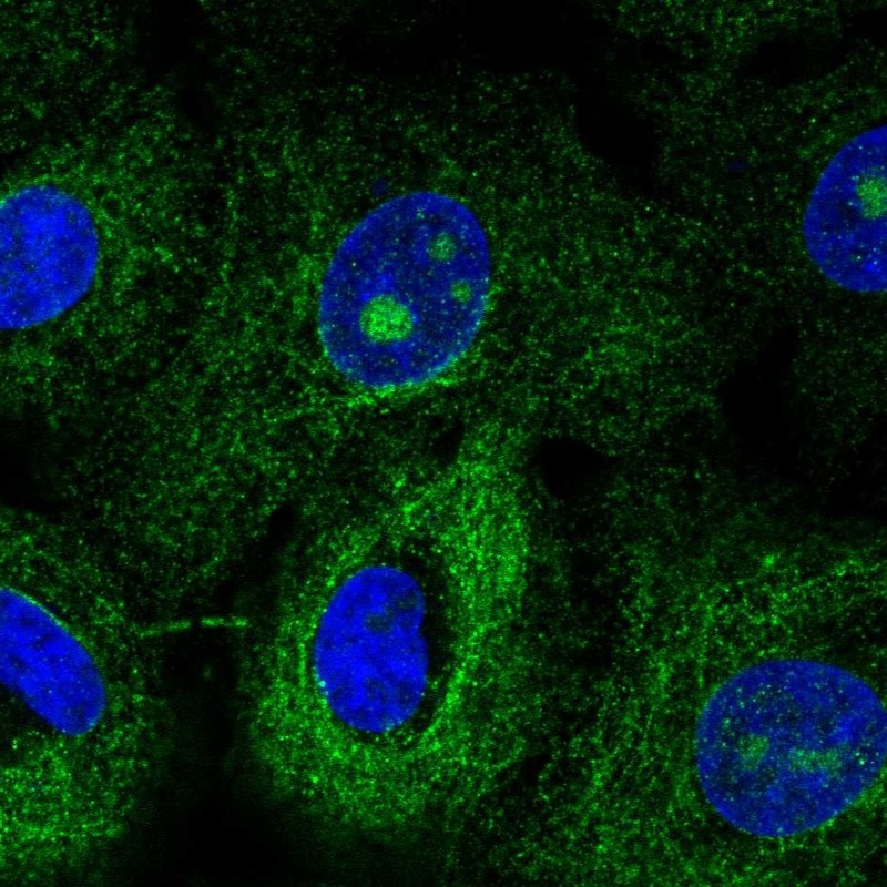
Figure 1. IF analysis of MTUS1 using anti-MTUS1 antibody (A04047-1). MTUS1 was detected in immunocytochemical section of U20S cell. Enzyme antigen retrieval was performed using IHC enzyme antigen retrieval reagent (AR0022) for 15 mins. The cells were blocked with 10% goat serum. And then incubated with 2microg/mL rabbit anti-MTUS1 Antibody (A04047-1) overnight at 4°C. Cy3 Conjugated Goat Anti-Rabbit IgG (BA1032) was used as secondary antibody at 1:100 dilution and incubated for 30 minutes at 37°C. The section was counterstained with DAPI. Visualize using a fluorescence microscope and filter sets appropriate for the label used.
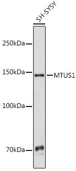
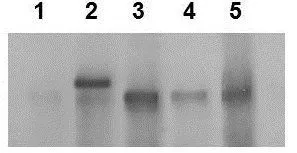
. Electrophoresis was performed on a 5-20% SDS-PAGE gel at 70V (Stacking gel) / 90V (Resolving gel) for 2-3 hours. The sample well of each lane was loaded with 50ug of sample under reducing conditions. Lane 1: human A549 whole cell lysates, Lane 2: human HepG2 whole cell lysates, Lane 3: human HEK293 whole cell lysates, Lane 4: human Caco-2 whole cell lysates, Lane 5: human Hela whole cell lysates. After Electrophoresis, proteins were transferred to a Nitrocellulose membrane at 150mA for 50-90 minutes. Blocked the membrane with 5% Non-fat Milk/ TBS for 1.5 hour at RT. The membrane was incubated with rabbit anti-MTUS1 antigen affinity purified polyclonal antibody (Catalog # A04047-1) at 0.5 microg/mL overnight at 4°C, then washed with TBS-0.1%Tween 3 times with 5 minutes each and probed with a goat anti-rabbit IgG-HRP secondary antibody at a dilution of 1:10000 for 1.5 hour at RT. The signal is developed using an Enhanced Chemiluminescent detection (ECL) kit (Catalog # EK1002) with Tanon 5200 system. A specific band was detected for MTUS1 at approximately 170KD. The expected band size for MTUS1 is at 141KD.")
Figure 1. IF analysis of MTUS1 using anti-MTUS1 antibody (A04047-1). MTUS1 was detected in immunocytochemical section of U20S cell. Enzyme antigen retrieval was performed using IHC enzyme antigen retrieval reagent (AR0022) for 15 mins. The cells were blocked with 10% goat serum. And then incubated with 2microg/mL rabbit anti-MTUS1 Antibody (A04047-1) overnight at 4°C. Cy3 Conjugated Goat Anti-Rabbit IgG (BA1032) was used as secondary antibody at 1:100 dilution and incubated for 30 minutes at 37°C. The section was counterstained with DAPI. Visualize using a fluorescence microscope and filter sets appropriate for the label used.
Anti-MTUS1 Antibody Picoband(r)
A04047-1-CARRIER-FREE
ApplicationsImmunoFluorescence, Western Blot, ELISA, ImmunoCytoChemistry
Product group Antibodies
ReactivityHuman, Mouse, Rat
TargetMTUS1
Overview
- SupplierBoster Bio
- Product NameAnti-MTUS1 Antibody Picoband(r)
- Delivery Days Customer9
- ApplicationsImmunoFluorescence, Western Blot, ELISA, ImmunoCytoChemistry
- CertificationResearch Use Only
- ClonalityPolyclonal
- Concentration500 ug/ml
- Gene ID57509
- Target nameMTUS1
- Target descriptionmicrotubule associated scaffold protein 1
- Target synonymsATBP, ATIP, ATIP3, ICIS, MP44, MTSG1, microtubule-associated tumor suppressor 1, AT2 receptor-binding protein, AT2 receptor-interacting protein, AT2R binding protein, angiotensin-II type 2 receptor-interacting protein, erythroid differentiation-related, microtubule associated tumor suppressor 1, mitochondrial tumor suppressor gene 1, transcription factor MTSG1
- HostRabbit
- IsotypeIgG
- Protein IDQ9ULD2
- Protein NameMicrotubule-associated tumor suppressor 1
- Scientific DescriptionBoster Bio Anti-MTUS1 Antibody Picoband® catalog # A04047-1. Tested in ELISA, IF, ICC, WB applications. This antibody reacts with Human, Mouse, Rat. The brand Picoband indicates this is a premium antibody that guarantees superior quality, high affinity, and strong signals with minimal background in Western blot applications. Only our best-performing antibodies are designated as Picoband, ensuring unmatched performance.
- ReactivityHuman, Mouse, Rat
- Storage Instruction-20°C,2°C to 8°C
- UNSPSC12352203
Related products
Product group Antibodies
MTUS1 AntibodyCSB-PA026004
ApplicationsELISA, ImmunoHistoChemistry
ReactivityHuman, Mouse, Rat
TargetMTUS1
- SizePrice
Product group Antibodies
Anti-MTUS1 AntibodyHPA069795
ApplicationsImmunoCytoChemistry
ReactivityHuman
TargetMTUS1
- SizePrice
Product group Antibodies

MTUS1 AntibodyLS-C401860
ApplicationsELISA, ImmunoHistoChemistry
ReactivityHuman, Mouse, Rat
TargetMTUS1
- SizePrice
Product group Antibodies
ApplicationsImmunoPrecipitation, Western Blot, ImmunoCytoChemistry, ImmunoHistoChemistry
ReactivityRat
TargetMTUS1
- SizePrice
Product group Antibodies
MTUS1 antibody, InternalGTX46731
ApplicationsWestern Blot, ImmunoHistoChemistry, ImmunoHistoChemistry Paraffin
ReactivityHuman
TargetMTUS1
- SizePrice
Product group Antibodies
Anti-MTUS1 (C-term) Antibody102-25021
ApplicationsWestern Blot
TargetMTUS1
- SizePrice
Product group Antibodies
References
MTUS1 Polyclonal AntibodyBS-6076R
ApplicationsImmunoFluorescence, Western Blot, ELISA, ImmunoCytoChemistry, ImmunoHistoChemistry, ImmunoHistoChemistry Frozen, ImmunoHistoChemistry Paraffin
ReactivityBovine, Canine, Chicken, Equine, Human, Mouse, Rabbit, Rat
TargetMTUS1
- SizePrice