


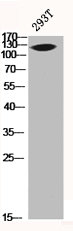
Figure 1. Western blot analysis of MUC1 using anti-MUC1 antibody (A00187-2). Electrophoresis was performed on a 5-20% SDS-PAGE gel at 70V (Stacking gel) / 90V (Resolving gel) for 2-3 hours. The sample well of each lane was loaded with 50ug of sample under reducing conditions. Lane 1: rat PC-12 whole cell lysates. After Electrophoresis, proteins were transferred to a Nitrocellulose membrane at 150mA for 50-90 minutes. Blocked the membrane with 5% Non-fat Milk/ TBS for 1.5 hour at RT. The membrane was incubated with rabbit anti-MUC1 antigen affinity purified polyclonal antibody (Catalog # A00187-2) at 0.5 microg/mL overnight at 4°C, then washed with TBS-0.1%Tween 3 times with 5 minutes each and probed with a goat anti-rabbit IgG-HRP secondary antibody at a dilution of 1:5000 for 1.5 hour at RT. The signal is developed using an Enhanced Chemiluminescent detection (ECL) kit (Catalog # EK1002) with Tanon 5200 system. A specific band was detected for MUC1 at approximately 122KD. The expected band size for MUC1 is at 122KD.
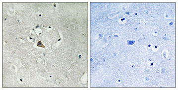
. MUC1 was detected in paraffin-embedded section of human rectal cancer tissue. Heat mediated antigen retrieval was performed in EDTA buffer (pH8.0, epitope retrieval solution). The tissue section was blocked with 10% goat serum. The tissue section was then incubated with 2microg/ml rabbit anti-MUC1 Antibody (A00187-2) overnight at 4°C. Biotinylated goat anti-rabbit IgG was used as secondary antibody and incubated for 30 minutes at 37°C. The tissue section was developed using Strepavidin-Biotin-Complex (SABC) (Catalog # SA1022) with DAB as the chromogen.")
. MUC1 was detected in paraffin-embedded section of human renal cancer tissue. Heat mediated antigen retrieval was performed in EDTA buffer (pH8.0, epitope retrieval solution). The tissue section was blocked with 10% goat serum. The tissue section was then incubated with 2microg/ml rabbit anti-MUC1 Antibody (A00187-2) overnight at 4°C. Biotinylated goat anti-rabbit IgG was used as secondary antibody and incubated for 30 minutes at 37°C. The tissue section was developed using Strepavidin-Biotin-Complex (SABC) (Catalog # SA1022) with DAB as the chromogen.")
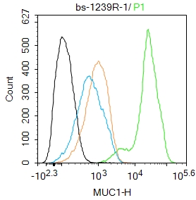
. Overlay histogram showing Hela cells stained with A00187-2 (Blue line). To facilitate intracellular staining, cells were fixed with 4% paraformaldehyde and permeabilized with permeabilization buffer. The cells were blocked with 10% normal goat serum. And then incubated with rabbit anti-MUC1 Antibody (A00187-2, 1microg/1x106 cells) for 30 min at 20°C. DyLight®488 conjugated goat anti-rabbit IgG (BA1127, 5-10microg/1x106 cells) was used as secondary antibody for 30 minutes at 20°C. Isotype control antibody (Green line) was rabbit IgG (1microg/1x106) used under the same conditions. Unlabelled sample without incubation with primary antibody and secondary antibody (Red line) was used as a blank control.")
Figure 1. Western blot analysis of MUC1 using anti-MUC1 antibody (A00187-2). Electrophoresis was performed on a 5-20% SDS-PAGE gel at 70V (Stacking gel) / 90V (Resolving gel) for 2-3 hours. The sample well of each lane was loaded with 50ug of sample under reducing conditions. Lane 1: rat PC-12 whole cell lysates. After Electrophoresis, proteins were transferred to a Nitrocellulose membrane at 150mA for 50-90 minutes. Blocked the membrane with 5% Non-fat Milk/ TBS for 1.5 hour at RT. The membrane was incubated with rabbit anti-MUC1 antigen affinity purified polyclonal antibody (Catalog # A00187-2) at 0.5 microg/mL overnight at 4°C, then washed with TBS-0.1%Tween 3 times with 5 minutes each and probed with a goat anti-rabbit IgG-HRP secondary antibody at a dilution of 1:5000 for 1.5 hour at RT. The signal is developed using an Enhanced Chemiluminescent detection (ECL) kit (Catalog # EK1002) with Tanon 5200 system. A specific band was detected for MUC1 at approximately 122KD. The expected band size for MUC1 is at 122KD.
Anti-MUC1 Antibody Picoband(r)
A00187-2-CARRIER-FREE
ApplicationsFlow Cytometry, Western Blot, ELISA, ImmunoHistoChemistry
Product group Antibodies
ReactivityHuman, Rat
TargetMUC1
Overview
- SupplierBoster Bio
- Product NameAnti-MUC1 Antibody Picoband(r)
- Delivery Days Customer9
- ApplicationsFlow Cytometry, Western Blot, ELISA, ImmunoHistoChemistry
- CertificationResearch Use Only
- ClonalityPolyclonal
- Concentration500 ug/ml
- Gene ID4582
- Target nameMUC1
- Target descriptionmucin 1, cell surface associated
- Target synonymsADMCKD, ADMCKD1, ADTKD2, CA 15-3, CD227, Ca15-3, EMA, H23AG, KL-6, MAM6, MCD, MCKD, MCKD1, MUC-1, MUC-1/SEC, MUC-1/X, MUC1/ZD, PEM, PEMT, PUM, mucin-1, H23 antigen, breast carcinoma-associated antigen DF3, cancer antigen 15-3, carcinoma-associated mucin, episialin, krebs von den Lungen-6, mucin 1, transmembrane, peanut-reactive urinary mucin, polymorphic epithelial mucin, tumor associated epithelial mucin, tumor-associated epithelial membrane antigen
- HostRabbit
- IsotypeIgG
- Protein IDP15941
- Protein NameMucin-1
- Scientific DescriptionBoster Bio Anti-MUC1 Antibody Picoband® catalog # A00187-2. Tested in ELISA, Flow Cytometry, IHC, WB applications. This antibody reacts with Human, Rat. The brand Picoband indicates this is a premium antibody that guarantees superior quality, high affinity, and strong signals with minimal background in Western blot applications. Only our best-performing antibodies are designated as Picoband, ensuring unmatched performance.
- ReactivityHuman, Rat
- Storage Instruction-20°C,2°C to 8°C
- UNSPSC12352203
Related products
Product group Antibodies

Anti-MUC1 [SM3]Ab00215-1.1
ApplicationsFlow Cytometry, ELISA, ImmunoHistoChemistry, ImmunoHistoChemistry Frozen, ImmunoHistoChemistry Paraffin
ReactivityHuman
TargetMUC1
- SizePrice
Product group Antibodies
Anti-MUC1 AntibodyA101263
ApplicationsELISA, ImmunoHistoChemistry
ReactivityHuman
- SizePrice
Product group Antibodies
Anti-MUC1 Antibody144-00333
ApplicationsWestern Blot
ReactivityHuman, Mouse
TargetMUC1
- SizePrice


Product group Antibodies
Anti-MUC1 AntibodyAMAB91533
ApplicationsImmunoHistoChemistry
ReactivityHuman
TargetMUC1
- SizePrice
Product group Antibodies
MUC1 Polyclonal AntibodyBS-1239R
ApplicationsELISA, ImmunoHistoChemistry, ImmunoHistoChemistry Paraffin
ReactivityCanine, Equine, Human, Mouse, Rat
TargetMUC1
- SizePrice
Product group Antibodies
MUC1 AntibodyCSB-PA003334
ApplicationsImmunoFluorescence, Western Blot, ELISA, ImmunoHistoChemistry
ReactivityHuman
TargetMUC1
- SizePrice
Product group Antibodies
Muc1 Polyclonal AntibodyCAC07111
ApplicationsImmunoFluorescence, ELISA, ImmunoHistoChemistry
TargetMUC1
- SizePrice
Product group Antibodies
MUC1 antibodyGTX100459
ApplicationsImmunoFluorescence, ImmunoPrecipitation, Western Blot, ImmunoCytoChemistry, ImmunoHistoChemistry, ImmunoHistoChemistry Paraffin
ReactivityHuman
TargetMUC1
- SizePrice