


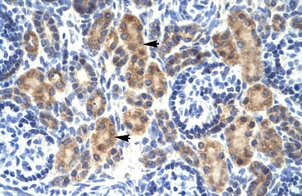
Immunohistochemical staining of human placenta shows moderate cytoplasmic positivity in trophoblastic cells.
Immunohistochemical staining of human placenta shows moderate cytoplasmic positivity in trophoblastic cells.
Anti-MYCBP Antibody
HPA041188
ApplicationsImmunoHistoChemistry
Product group Antibodies
ReactivityHuman
TargetMYCBP
Overview
- SupplierAtlas Antibodies
- Product NameAnti-MYCBP Antibody
- Delivery Days Customer4
- ApplicationsImmunoHistoChemistry
- CertificationResearch Use Only
- ClonalityPolyclonal
- ConjugateUnconjugated
- Gene ID26292
- Target nameMYCBP
- Target descriptionMYC binding protein
- Target synonymsAMY-1, C-Myc-binding protein, associate of myc-1, c-myc binding protein
- HostRabbit
- IsotypeIgG
- Protein IDQ99417
- Protein Namec-Myc-binding protein
- Scientific DescriptionRecombinant Protein Epitope Signature Tag (PrEST) antigen sequence
- ReactivityHuman
- Storage Instruction-20°C,2°C to 8°C
- UNSPSC41116161
Datasheet
MSDS
Related products
Product group Antibodies
MYCBP AntibodyCSB-PA008741
ApplicationsELISA, ImmunoHistoChemistry
ReactivityHuman, Mouse
TargetMYCBP
- SizePrice

Product group Antibodies
Anti-MYCBP Antibody Picoband(r)A06216-2-CARRIER-FREE
ApplicationsFlow Cytometry, Western Blot, ELISA, ImmunoHistoChemistry
ReactivityHuman, Mouse, Rat
TargetMYCBP
- SizePrice
Product group Antibodies

MYCBP Antibody (aa2-103, HRP)LS-C371968
ApplicationsELISA, ImmunoHistoChemistry, ImmunoHistoChemistry Paraffin
ReactivityHuman
TargetMYCBP
- SizePrice
Product group Antibodies
MYCBP Monoclonal AntibodyCAC13668
ApplicationsELISA, ImmunoHistoChemistry
TargetMYCBP
- SizePrice
Product group Antibodies
MYCBP antibody, InternalGTX49061
ApplicationsWestern Blot, ImmunoHistoChemistry, ImmunoHistoChemistry Paraffin
ReactivityHuman
TargetMYCBP
- SizePrice
Product group Antibodies
Anti-MYCBP Antibody144-61391
ApplicationsWestern Blot
ReactivityHuman
TargetMYCBP
- SizePrice