

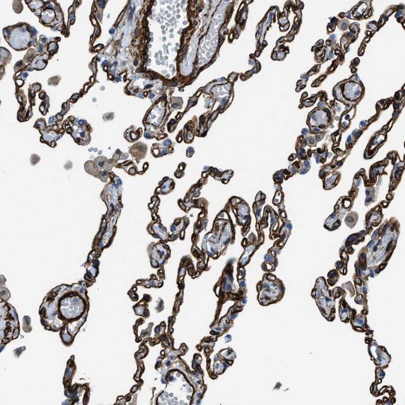
Immunohistochemical staining of human lung shows strong membranous positivity in pneumocytes.
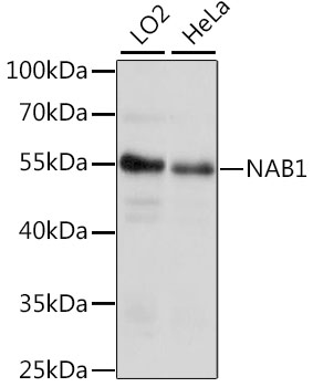
 and NAB1 over-expression lysate (Co-expressed with a C-terminal myc-DDK tag (~3.1 kDa) in mammalian HEK293T cells, LY401805).")
Immunohistochemical staining of human lung shows strong membranous positivity in pneumocytes.
Anti-NAB1 Antibody
HPA002738
ApplicationsWestern Blot, ImmunoCytoChemistry, ImmunoHistoChemistry
Product group Antibodies
ReactivityHuman
TargetNAB1
Overview
- SupplierAtlas Antibodies
- Product NameAnti-NAB1 Antibody
- Delivery Days Customer4
- ApplicationsWestern Blot, ImmunoCytoChemistry, ImmunoHistoChemistry
- CertificationResearch Use Only
- ClonalityPolyclonal
- ConjugateUnconjugated
- Gene ID4664
- Target nameNAB1
- Target descriptionNGFI-A binding protein 1
- Target synonymsNGFI-A-binding protein 1, EGR-1-binding protein 1, EGR1 binding protein 1, transcriptional regulatory protein p54
- HostRabbit
- IsotypeIgG
- Protein IDQ13506
- Protein NameNGFI-A-binding protein 1
- Scientific DescriptionRecombinant Protein Epitope Signature Tag (PrEST) antigen sequence
- ReactivityHuman
- Storage Instruction-20°C,2°C to 8°C
- UNSPSC41116161
Datasheet
MSDS
Related products
Product group Antibodies
Anti-NAB1 Antibody144-63875
ApplicationsWestern Blot
ReactivityHuman
TargetNAB1
- SizePrice

Product group Antibodies
Anti-NAB1 Antibody Picoband(r)A06669-1-CARRIER-FREE
ApplicationsImmunoFluorescence, Western Blot, ELISA, ImmunoCytoChemistry
ReactivityHuman, Rat
TargetNAB1
- SizePrice
Product group Antibodies

NAB1 Antibody (aa410-460)LS-C289782
ApplicationsImmunoPrecipitation
ReactivityHuman
TargetNAB1
- SizePrice
Product group Antibodies
NAB1 antibodyGTX112354
ApplicationsImmunoFluorescence, Western Blot, ImmunoCytoChemistry
ReactivityHuman
TargetNAB1
- SizePrice