


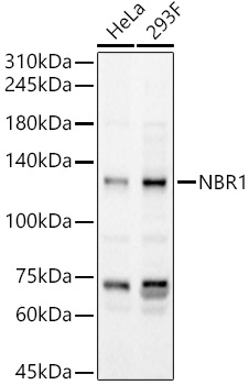
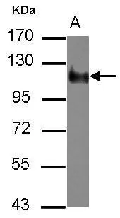
Figure 1. Western blot analysis of NBR1 using anti-NBR1 antibody (A03534-1). Electrophoresis was performed on a 5-20% SDS-PAGE gel at 70V (Stacking gel) / 90V (Resolving gel) for 2-3 hours. The sample well of each lane was loaded with 30 ug of sample under reducing conditions. Lane 1: human Hela whole cell lysates, Lane 2: human HepG2 whole cell lysates, Lane 3: human K562 whole cell lysates, Lane 4: monkey COS-7 whole cell lysates. After electrophoresis, proteins were transferred to a nitrocellulose membrane at 150 mA for 50-90 minutes. Blocked the membrane with 5% non-fat milk/TBS for 1.5 hour at RT. The membrane was incubated with rabbit anti-NBR1 antigen affinity purified polyclonal antibody (Catalog # A03534-1) at 0.5 microg/mL overnight at 4°C, then washed with TBS-0.1%Tween 3 times with 5 minutes each and probed with a goat anti-rabbit IgG-HRP secondary antibody at a dilution of 1:5000 for 1.5 hour at RT. The signal is developed using an Enhanced Chemiluminescent detection (ECL) kit (Catalog # EK1002) with Tanon 5200 system. A specific band was detected for NBR1 at approximately 140 kDa. The expected band size for NBR1 is at 107,140 kDa.
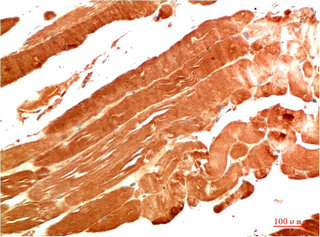
 and anti-Beta Tubulin antibody (M01857-3). NBR1 was detected in immunocytochemical section of A549 cell. Enzyme antigen retrieval was performed using IHC enzyme antigen retrieval reagent (AR0022) for 15 mins. The cells were blocked with 10% goat serum. And then incubated with 5 microg/mL rabbit anti-NBR1 Antibody (A03534-1) and mouse anti-Beta Tubulin antibody (M01857-3) overnight at 4°C. Cy3 Conjugated Goat Anti-Rabbit IgG (BA1032) and DyLight®488 Conjugated Goat Anti-Mouse IgG (BA1126) were used as secondary antibody at 1:500 dilution and incubated for 30 minutes at 37°C. Visualize using a fluorescence microscope and filter sets appropriate for the label used.")
. Overlay histogram showing MCF-7 cells stained with A03534-1 (Blue line). To facilitate intracellular staining, cells were fixed with 4% paraformaldehyde and permeabilized with permeabilization buffer. The cells were blocked with 10% normal goat serum. And then incubated with rabbit anti-NBR1 Antibody (A03534-1, 1 microg/1x106 cells) for 30 min at 20°C. DyLight®488 conjugated goat anti-rabbit IgG (BA1127, 5-10 microg/1x106 cells) was used as secondary antibody for 30 minutes at 20°C. Isotype control antibody (Green line) was rabbit IgG (1 microg/1x106) used under the same conditions. Unlabelled sample (Red line) was also used as a control.")
Figure 1. Western blot analysis of NBR1 using anti-NBR1 antibody (A03534-1). Electrophoresis was performed on a 5-20% SDS-PAGE gel at 70V (Stacking gel) / 90V (Resolving gel) for 2-3 hours. The sample well of each lane was loaded with 30 ug of sample under reducing conditions. Lane 1: human Hela whole cell lysates, Lane 2: human HepG2 whole cell lysates, Lane 3: human K562 whole cell lysates, Lane 4: monkey COS-7 whole cell lysates. After electrophoresis, proteins were transferred to a nitrocellulose membrane at 150 mA for 50-90 minutes. Blocked the membrane with 5% non-fat milk/TBS for 1.5 hour at RT. The membrane was incubated with rabbit anti-NBR1 antigen affinity purified polyclonal antibody (Catalog # A03534-1) at 0.5 microg/mL overnight at 4°C, then washed with TBS-0.1%Tween 3 times with 5 minutes each and probed with a goat anti-rabbit IgG-HRP secondary antibody at a dilution of 1:5000 for 1.5 hour at RT. The signal is developed using an Enhanced Chemiluminescent detection (ECL) kit (Catalog # EK1002) with Tanon 5200 system. A specific band was detected for NBR1 at approximately 140 kDa. The expected band size for NBR1 is at 107,140 kDa.
Anti-NBR1 Antibody Picoband(r)
A03534-1-CARRIER-FREE
ApplicationsFlow Cytometry, ImmunoFluorescence, Western Blot, ELISA, ImmunoCytoChemistry
Product group Antibodies
ReactivityHuman, Monkey
TargetNBR1
Overview
- SupplierBoster Bio
- Product NameAnti-NBR1 Antibody Picoband(r)
- Delivery Days Customer9
- ApplicationsFlow Cytometry, ImmunoFluorescence, Western Blot, ELISA, ImmunoCytoChemistry
- CertificationResearch Use Only
- ClonalityPolyclonal
- Concentration500 ug/ml
- Gene ID4077
- Target nameNBR1
- Target descriptionNBR1 autophagy cargo receptor
- Target synonyms1A1-3B, IAI3B, M17S2, MIG19, next to BRCA1 gene 1 protein, B-box protein, cell migration-inducing gene 19 protein, membrane component, chromosome 17, surface marker 2 (ovarian carcinoma antigen CA125), migration-inducing protein 19, neighbor of BRCA1 gene 1
- HostRabbit
- IsotypeIgG
- Protein IDQ14596
- Protein NameNext to BRCA1 gene 1 protein
- Scientific DescriptionBoster Bio Anti-NBR1 Antibody Picoband® catalog # A03534-1. Tested in ELISA, IF, ICC, WB, Flow Cytometry applications. This antibody reacts with Human, Monkey. The brand Picoband indicates this is a premium antibody that guarantees superior quality, high affinity, and strong signals with minimal background in Western blot applications. Only our best-performing antibodies are designated as Picoband, ensuring unmatched performance.
- ReactivityHuman, Monkey
- Storage Instruction-20°C,2°C to 8°C
- UNSPSC12352203
Related products
Product group Antibodies
Anti-NBR1 AntibodyA16697
ApplicationsImmunoFluorescence, ImmunoPrecipitation, Western Blot, ImmunoCytoChemistry, ImmunoHistoChemistry
ReactivityHuman, Mouse, Rat
- SizePrice
Product group Antibodies
Anti-NBR1 Antibody144-65754
ApplicationsImmunoFluorescence, ImmunoPrecipitation, Western Blot, ImmunoHistoChemistry
ReactivityHuman, Monkey, Mouse, Rat
TargetNBR1
- SizePrice
Product group Antibodies

NBR1 Antibody (clone 4C6)LS-C768427
ApplicationsImmunoFluorescence, ImmunoHistoChemistry, ImmunoHistoChemistry Paraffin
ReactivityHuman, Mouse, Rat
TargetNBR1
- SizePrice
Product group Antibodies
NBR1 Monoclonal AntibodyCSB-MA406807
ApplicationsELISA, ImmunoHistoChemistry
ReactivityHuman, Mouse, Rat
TargetNBR1
- SizePrice

Product group Antibodies
Anti-NBR1 AntibodyHPA022944
ApplicationsImmunoCytoChemistry
ReactivityHuman
TargetNBR1
- SizePrice
Product group Antibodies
NBR1 antibodyGTX114539
ApplicationsWestern Blot, ImmunoHistoChemistry, ImmunoHistoChemistry Paraffin
ReactivityHuman, Mouse
TargetNBR1
- SizePrice