


Immunofluorescent staining of human cell line MCF7 shows localization to nucleoplasm.

Immunofluorescent staining of human cell line MCF7 shows localization to nucleoplasm.
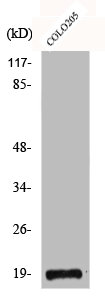
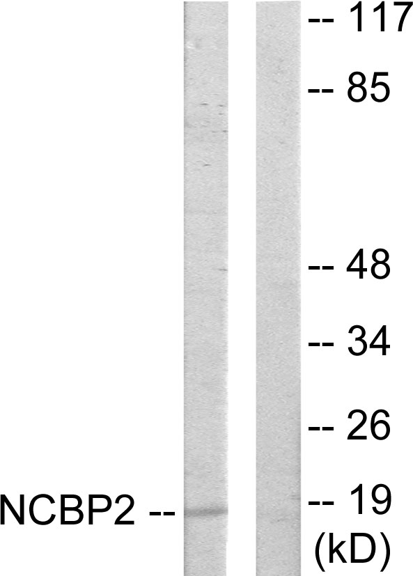
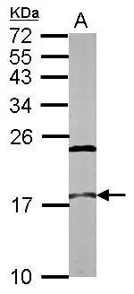
Anti-NCBP2 Antibody
HPA062483
ApplicationsWestern Blot, ImmunoCytoChemistry
Product group Antibodies
ReactivityHuman
TargetNCBP2
Overview
- SupplierAtlas Antibodies
- Product NameAnti-NCBP2 Antibody
- Delivery Days Customer4
- ApplicationsWestern Blot, ImmunoCytoChemistry
- CertificationResearch Use Only
- ClonalityPolyclonal
- ConjugateUnconjugated
- Gene ID22916
- Target nameNCBP2
- Target descriptionnuclear cap binding protein subunit 2
- Target synonymsCBC2, CBP20, NIP1, PIG55, nuclear cap-binding protein subunit 2, 20 kDa nuclear cap-binding protein, NCBP 20 kDa subunit, NCBP interacting protein 1, cell proliferation-inducing gene 55 protein, nuclear cap binding protein subunit 2, 20kD, nuclear cap binding protein subunit 2, 20kDa
- HostRabbit
- IsotypeIgG
- Protein IDP52298
- Protein NameNuclear cap-binding protein subunit 2
- Scientific DescriptionRecombinant Protein Epitope Signature Tag (PrEST) antigen sequence
- ReactivityHuman
- Storage Instruction-20°C,2°C to 8°C
- UNSPSC41116161
Datasheet
MSDS
Related products
Product group Antibodies
Anti-NCBP2 Antibody144-07293
ApplicationsImmunoFluorescence, Western Blot, ImmunoHistoChemistry
ReactivityHuman, Mouse, Rat
TargetNCBP2
- SizePrice
Product group Antibodies
NCBP2 AntibodyCSB-PA001369
ApplicationsImmunoFluorescence, Western Blot, ELISA, ImmunoHistoChemistry
ReactivityHuman, Mouse
TargetNCBP2
- SizePrice
Product group Antibodies
Anti-NCBP2 AntibodyA99025
ApplicationsWestern Blot, ELISA
ReactivityHuman, Mouse
- SizePrice
Product group Antibodies

NCBP2 / CBP20 Antibody (N-Terminus)LS-C368657
ApplicationsImmunoFluorescence, Western Blot, ImmunoCytoChemistry, ImmunoHistoChemistry, ImmunoHistoChemistry Paraffin
ReactivityBovine, Human, Monkey, Mouse, Rat
TargetNCBP2
- SizePrice
Product group Antibodies
NCBP2 antibody [N1C3]GTX115648
ApplicationsWestern Blot
ReactivityHuman, Mouse
TargetNCBP2
- SizePrice

Product group Antibodies
Anti-CBP20/NCBP2 Antibody Picoband(r)A08031-2-CARRIER-FREE
ApplicationsFlow Cytometry, ImmunoFluorescence, Western Blot, ELISA, ImmunoCytoChemistry, ImmunoHistoChemistry
ReactivityHuman, Mouse, Rat
TargetNCBP2
- SizePrice