

Anti-NDUFA1 Antibody
A28656
ApplicationsWestern Blot
Product group Antibodies
ReactivityHuman, Mouse, Rat
Overview
- SupplierAntibodies.com
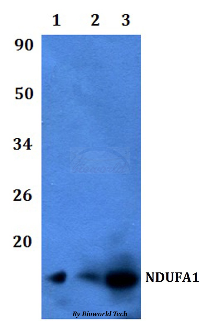
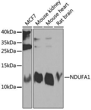
- Product NameAnti-NDUFA1 Antibody
- Delivery Days Customer7
- ApplicationsWestern Blot
- CertificationResearch Use Only
- ClonalityPolyclonal
- ConjugateUnconjugated
- Estimated Purity>95%
- HostRabbit
- Scientific DescriptionRabbit polyclonal antibody to NDUFA1
- ReactivityHuman, Mouse, Rat
- UNSPSC12352203
Related products

Product group Antibodies
Anti-NDUFA1 Antibody Picoband(r)A08224-CARRIER-FREE
ApplicationsWestern Blot, ELISA, ImmunoHistoChemistry
ReactivityHuman, Mouse, Rat
TargetNDUFA1
- SizePrice
Product group Antibodies
Anti-NDUFA1 Antibody144-08326
ApplicationsWestern Blot
ReactivityHuman, Mouse, Rat
TargetNDUFA1
- SizePrice
Product group Antibodies
NDUFA1 Polyclonal AntibodyBS-3956R
ApplicationsImmunoFluorescence, ELISA, ImmunoCytoChemistry, ImmunoHistoChemistry, ImmunoHistoChemistry Frozen, ImmunoHistoChemistry Paraffin
ReactivityCanine, Equine, Human, Mouse, Rat
TargetNDUFA1
- SizePrice
Product group Antibodies
NDUFA1 AntibodyCSB-PA01665A0RB
ApplicationsImmunoFluorescence, ELISA, ImmunoHistoChemistry
ReactivityHuman
TargetNDUFA1
- SizePrice
Product group Antibodies

NDUFA1 AntibodyLS-C401871
ApplicationsELISA, ImmunoHistoChemistry
ReactivityHuman
TargetNDUFA1
- SizePrice

Product group Antibodies
Anti-NDUFA1 AntibodyHPA029768
ApplicationsImmunoCytoChemistry
ReactivityHuman
TargetNDUFA1
- SizePrice
Product group Antibodies
NDUFA1 antibodyGTX32740
ApplicationsWestern Blot
ReactivityHuman, Mouse, Rat
TargetNDUFA1
- SizePrice
Product group Antibodies
Anti-NDUFA1 AntibodyCAB8326
ApplicationsWestern Blot, ELISA
ReactivityHuman
TargetNDUFA1
- SizePrice