


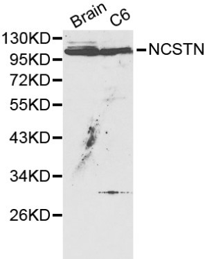
Figure 1. Western blot analysis of Nicastrin/NCSTN using anti-Nicastrin/NCSTN antibody (A03144-1). Electrophoresis was performed on a 5-20% SDS-PAGE gel at 70V (Stacking gel) / 90V (Resolving gel) for 2-3 hours. The sample well of each lane was loaded with 30 ug of sample under reducing conditions. Lane 1: human THP-1 whole cell lysates, Lane 2: human Hela whole cell lysates, Lane 3: human HL-60 whole cell lysates, Lane 4: human HepG2 whole cell lysates, Lane 5: rat lung tissue lysates, Lane 6: rat pancreas tissue lysates, Lane 7: mouse lung tissue lysates, Lane 8: mouse pancreas tissue lysates. After electrophoresis, proteins were transferred to a nitrocellulose membrane at 150 mA for 50-90 minutes. Blocked the membrane with 5% non-fat milk/TBS for 1.5 hour at RT. The membrane was incubated with rabbit anti-Nicastrin/NCSTN antigen affinity purified polyclonal antibody (Catalog # A03144-1) at 0.5 microg/mL overnight at 4°C, then washed with TBS-0.1%Tween 3 times with 5 minutes each and probed with a goat anti-rabbit IgG-HRP secondary antibody at a dilution of 1:5000 for 1.5 hour at RT. The signal is developed using an Enhanced Chemiluminescent detection (ECL) kit (Catalog # EK1002) with Tanon 5200 system. A specific band was detected for Nicastrin/NCSTN at approximately 120 kDa. The expected band size for Nicastrin/NCSTN is at 120 kDa.
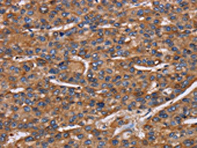
. Nicastrin/NCSTN was detected in an immunocytochemical section of SiHa cells. Enzyme antigen retrieval was performed using IHC enzyme antigen retrieval reagent (AR0022) for 15 mins. The cells were blocked with 10% goat serum. And then incubated with 5 microg/mL rabbit anti-Nicastrin/NCSTN Antibody (A03144-1) overnight at 4°C. DyLight®594 Conjugated Goat Anti-Rabbit IgG (BA1142) was used as secondary antibody at 1:100 dilution and incubated for 30 minutes at 37°C. The section was counterstained with DAPI. Visualize using a fluorescence microscope and filter sets appropriate for the label used.")
Figure 1. Western blot analysis of Nicastrin/NCSTN using anti-Nicastrin/NCSTN antibody (A03144-1). Electrophoresis was performed on a 5-20% SDS-PAGE gel at 70V (Stacking gel) / 90V (Resolving gel) for 2-3 hours. The sample well of each lane was loaded with 30 ug of sample under reducing conditions. Lane 1: human THP-1 whole cell lysates, Lane 2: human Hela whole cell lysates, Lane 3: human HL-60 whole cell lysates, Lane 4: human HepG2 whole cell lysates, Lane 5: rat lung tissue lysates, Lane 6: rat pancreas tissue lysates, Lane 7: mouse lung tissue lysates, Lane 8: mouse pancreas tissue lysates. After electrophoresis, proteins were transferred to a nitrocellulose membrane at 150 mA for 50-90 minutes. Blocked the membrane with 5% non-fat milk/TBS for 1.5 hour at RT. The membrane was incubated with rabbit anti-Nicastrin/NCSTN antigen affinity purified polyclonal antibody (Catalog # A03144-1) at 0.5 microg/mL overnight at 4°C, then washed with TBS-0.1%Tween 3 times with 5 minutes each and probed with a goat anti-rabbit IgG-HRP secondary antibody at a dilution of 1:5000 for 1.5 hour at RT. The signal is developed using an Enhanced Chemiluminescent detection (ECL) kit (Catalog # EK1002) with Tanon 5200 system. A specific band was detected for Nicastrin/NCSTN at approximately 120 kDa. The expected band size for Nicastrin/NCSTN is at 120 kDa.
Anti-Nicastrin/NCSTN Antibody Picoband(r)
A03144-1-CARRIER-FREE
ApplicationsImmunoFluorescence, Western Blot, ELISA, ImmunoCytoChemistry
Product group Antibodies
ReactivityHuman, Mouse, Rat
TargetNCSTN
Overview
- SupplierBoster Bio
- Product NameAnti-Nicastrin/NCSTN Antibody Picoband(r)
- Delivery Days Customer9
- ApplicationsImmunoFluorescence, Western Blot, ELISA, ImmunoCytoChemistry
- CertificationResearch Use Only
- ClonalityPolyclonal
- Concentration500 ug/ml
- Gene ID23385
- Target nameNCSTN
- Target descriptionnicastrin
- Target synonymsATAG1874, nicastrin, anterior pharynx-defective 2
- HostRabbit
- IsotypeIgG
- Protein IDQ92542
- Protein NameNicastrin
- Scientific DescriptionBoster Bio Anti-heavy chain Nicastrin/NCSTN Antibody Picoband® catalog # A03144-1. Tested in ELISA, IF, ICC, WB applications. This antibody reacts with Human, Mouse, Rat. The brand Picoband indicates this is a premium antibody that guarantees superior quality, high affinity, and strong signals with minimal background in Western blot applications. Only our best-performing antibodies are designated as Picoband, ensuring unmatched performance.
- ReactivityHuman, Mouse, Rat
- Storage Instruction-20°C,2°C to 8°C
- UNSPSC12352203
Related products
Product group Antibodies
Anti-NCSTN AntibodyA29354
ApplicationsWestern Blot, ImmunoHistoChemistry
ReactivityHuman, Mouse, Rat
- SizePrice
Product group Antibodies
Anti-NCSTN Antibody144-60683
ApplicationsWestern Blot
ReactivityHuman, Mouse, Rat
TargetNCSTN
- SizePrice
Product group Antibodies
References
Nicastrin Polyclonal AntibodyBS-6058R
ApplicationsImmunoFluorescence, Western Blot, ELISA, ImmunoCytoChemistry, ImmunoHistoChemistry, ImmunoHistoChemistry Frozen, ImmunoHistoChemistry Paraffin
ReactivityBovine, Chicken, Equine, Human, Mouse, Porcine, Rat
TargetNCSTN
- SizePrice
Product group Antibodies
ApplicationsImmunoPrecipitation, Western Blot, ImmunoCytoChemistry, ImmunoHistoChemistry
ReactivityRat
TargetNCSTN
- SizePrice
Product group Antibodies
NCSTN AntibodyCSB-PA214212
ApplicationsWestern Blot, ELISA, ImmunoHistoChemistry
ReactivityHuman, Mouse, Rat
TargetNCSTN
- SizePrice
Product group Antibodies

NCSTN / Nicastrin AntibodyLS-C401888
ApplicationsWestern Blot, ELISA, ImmunoHistoChemistry
ReactivityHuman, Mouse, Rat
TargetNCSTN
- SizePrice
Product group Antibodies
Nicastrin antibodyGTX23444
ApplicationsWestern Blot
ReactivityHuman, Mouse, Rat
TargetNCSTN
- SizePrice

Product group Antibodies
Anti-NCSTN AntibodyHPA054846
ApplicationsImmunoHistoChemistry
ReactivityHuman
TargetNCSTN
- SizePrice
Product group Antibodies
Anti-NCSTN AntibodyCAB0128
ApplicationsImmunoFluorescence, Western Blot, ELISA, ImmunoCytoChemistry
ReactivityHuman
TargetNCSTN
- SizePrice