

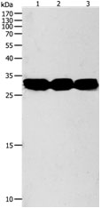

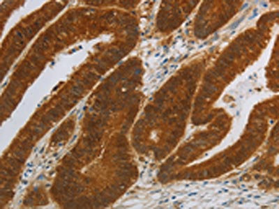
Anti-NIT2 Antibody
A48501
ApplicationsWestern Blot, ELISA, ImmunoHistoChemistry
Product group Antibodies
ReactivityHuman, Mouse
Overview
- SupplierAntibodies.com
- Product NameAnti-NIT2 Antibody
- Delivery Days Customer7
- ApplicationsWestern Blot, ELISA, ImmunoHistoChemistry
- CertificationResearch Use Only
- ClonalityPolyclonal
- ConjugateUnconjugated
- HostRabbit
- Scientific DescriptionRabbit polyclonal antibody to NIT2
- ReactivityHuman, Mouse
- UNSPSC12352203
Related products
Product group Antibodies
NIT2 AntibodyCSB-PA187627
ApplicationsWestern Blot, ELISA, ImmunoHistoChemistry
ReactivityHuman, Mouse
TargetNIT2
- SizePrice

Product group Antibodies
Anti-NIT2 Antibody Picoband(r)A05891-3-CARRIER-FREE
ApplicationsFlow Cytometry, Western Blot, ELISA, ImmunoHistoChemistry
ReactivityHuman, Mouse, Rat
TargetNIT2
- SizePrice

Product group Antibodies
Anti-NIT2 AntibodyHPA036999
ApplicationsWestern Blot, ImmunoCytoChemistry, ImmunoHistoChemistry
ReactivityHuman, Mouse, Rat
TargetNIT2
- SizePrice
Product group Antibodies

NIT2 AntibodyLS-C405959
ApplicationsWestern Blot, ELISA, ImmunoHistoChemistry
ReactivityHuman, Mouse
TargetNIT2
- SizePrice
![FACS analysis of HEK293T cells transfected with either NIT2 plasmid(Red) or empty vector control plasmid(Blue) using GTX84036 NIT2 antibody [2B9].](https://www.genetex.com/upload/website/prouct_img/normal/GTX84036/GTX84036_281_FACS_w_23061420_986.webp)
Product group Antibodies
NIT2 antibody [2B9]GTX84036
ApplicationsFlow Cytometry, ImmunoFluorescence, Western Blot, ImmunoCytoChemistry, ImmunoHistoChemistry, ImmunoHistoChemistry Paraffin
ReactivityHuman
TargetNIT2
- SizePrice
Product group Antibodies
NIT2 Polyclonal AntibodyCAC14456
ApplicationsWestern Blot, ELISA
ReactivityMouse
TargetNIT2
- SizePrice