


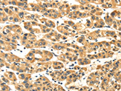
Immunohistochemical staining of human testis shows strong cytoplasmic positivity in subset of cells in seminiferous ducts.
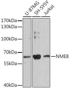
 and NME8 over-expression lysate (Co-expressed with a C-terminal myc-DDK tag (~3.1 kDa) in mammalian HEK293T cells, LY413869).")
Immunohistochemical staining of human testis shows strong cytoplasmic positivity in subset of cells in seminiferous ducts.
Anti-NME8 Antibody
HPA019259
ApplicationsWestern Blot, ImmunoHistoChemistry
Product group Antibodies
ReactivityHuman
TargetNME8
Overview
- SupplierAtlas Antibodies
- Product NameAnti-NME8 Antibody
- Delivery Days Customer4
- ApplicationsWestern Blot, ImmunoHistoChemistry
- CertificationResearch Use Only
- ClonalityPolyclonal
- ConjugateUnconjugated
- Gene ID51314
- Target nameNME8
- Target descriptionNME/NM23 family member 8
- Target synonymsCILD6, DNAI8, HEL-S-99, NM23-H8, SPTRX2, TXNDC3, sptrx-2, thioredoxin domain-containing protein 3, 3'-5' exonuclease NME8, epididymis secretory protein Li 99, sperm-specific thioredoxin 2, spermatid-specific thioredoxin-2, thioredoxin domain containing 3 (spermatozoa)
- HostRabbit
- IsotypeIgG
- Protein IDQ8N427
- Protein NameThioredoxin domain-containing protein 3
- Scientific DescriptionRecombinant Protein Epitope Signature Tag (PrEST) antigen sequence
- ReactivityHuman
- Storage Instruction-20°C,2°C to 8°C
- UNSPSC41116161
Datasheet
MSDS
Related products
Product group Antibodies
Anti-NME8 AntibodyA93271
ApplicationsWestern Blot
ReactivityHuman, Mouse
- SizePrice

Product group Antibodies
Anti-TXNDC3/NME8 Antibody Picoband(r)A08723-1-CARRIER-FREE
ApplicationsFlow Cytometry, Western Blot, ELISA
ReactivityHuman, Rat
TargetNME8
- SizePrice
Product group Antibodies
Anti-TXNDC3 Antibody107-10142
ApplicationsWestern Blot, ImmunoHistoChemistry, ImmunoHistoChemistry Paraffin
ReactivityHuman
TargetNME8
- SizePrice
Product group Antibodies

TXNDC3 AntibodyLS-C768565
ApplicationsELISA, ImmunoHistoChemistry
ReactivityHuman
TargetNME8
- SizePrice
Product group Antibodies
NME8 AntibodyCSB-PA109162
ApplicationsELISA, ImmunoHistoChemistry
ReactivityHuman
TargetNME8
- SizePrice


Product group Antibodies
Anti-NME8 AntibodyHPA029572
ApplicationsImmunoCytoChemistry
ReactivityHuman
TargetNME8
- SizePrice
Product group Antibodies
TXNDC3 antibody [N3C3]GTX111512
ApplicationsWestern Blot, ImmunoHistoChemistry, ImmunoHistoChemistry Paraffin
ReactivityHuman
TargetNME8
- SizePrice