


Immunofluorescent staining of human cell line U-2 OS shows localization to nucleus.
 shows similar pattern to independent antibody HPA043464 (B).")
Immunofluorescent staining of human cell line U-2 OS shows localization to nucleus.
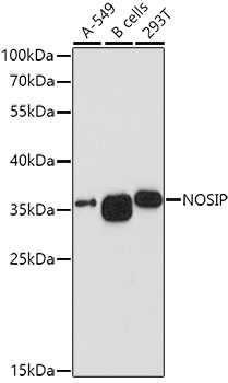
Anti-NOSIP Antibody
HPA062132
ApplicationsWestern Blot, ImmunoCytoChemistry
Product group Antibodies
ReactivityHuman
TargetNOSIP
Overview
- SupplierAtlas Antibodies
- Product NameAnti-NOSIP Antibody
- Delivery Days Customer4
- ApplicationsWestern Blot, ImmunoCytoChemistry
- CertificationResearch Use Only
- ClonalityPolyclonal
- ConjugateUnconjugated
- Gene ID51070
- Target nameNOSIP
- Target descriptionnitric oxide synthase interacting protein
- Target synonymsCGI-25, nitric oxide synthase-interacting protein, E3 ubiquitin-protein ligase NOSIP, RING-type E3 ubiquitin transferase NOSIP, eNOS-interacting protein
- HostRabbit
- IsotypeIgG
- Protein IDQ9Y314
- Protein NameNitric oxide synthase-interacting protein
- Scientific DescriptionRecombinant Protein Epitope Signature Tag (PrEST) antigen sequence
- ReactivityHuman
- Storage Instruction-20°C,2°C to 8°C
- UNSPSC41116161
Datasheet
MSDS
Related products

Product group Antibodies
Anti-NOSIP Antibody Picoband(r)A07558-2-CARRIER-FREE
ApplicationsFlow Cytometry, ImmunoFluorescence, Western Blot, ELISA, ImmunoCytoChemistry
ReactivityHuman
TargetNOSIP
- SizePrice
Product group Antibodies
Anti-NOSIP Antibody144-10024
ApplicationsWestern Blot
ReactivityHuman
TargetNOSIP
- SizePrice
Product group Antibodies
NOSIP AntibodyCSB-PA897477XA01HU
ApplicationsWestern Blot, ELISA
ReactivityHuman
TargetNOSIP
- SizePrice
Product group Antibodies
ApplicationsWestern Blot, ImmunoHistoChemistry
TargetNOSIP
- SizePrice
Product group Antibodies

NOSIP AntibodyLS-C496733
ApplicationsWestern Blot
ReactivityHuman
TargetNOSIP
- SizePrice

Product group Antibodies
Anti-NOSIP AntibodyHPA043464
ApplicationsWestern Blot, ImmunoCytoChemistry, ImmunoHistoChemistry
ReactivityHuman
TargetNOSIP
- SizePrice
Product group Antibodies
Anti-NOSIP AntibodyHPA043464
ApplicationsWestern Blot, ImmunoCytoChemistry, ImmunoHistoChemistry
ReactivityHuman
TargetNOSIP
- SizePrice