


Immunohistochemical staining of human cerebral cortex shows strong cytoplasmic positivity in neuronal cells.
Immunohistochemical staining of human cerebral cortex shows strong cytoplasmic positivity in neuronal cells.
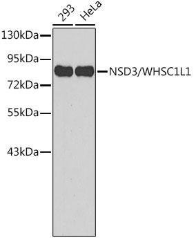
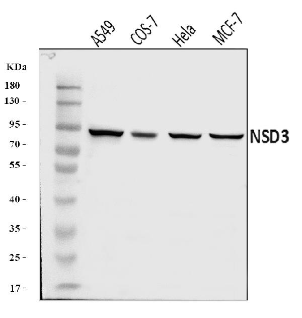
Anti-NSD3 Antibody
HPA005659
ApplicationsImmunoHistoChemistry
Product group Antibodies
ReactivityHuman
TargetNSD3
Overview
- SupplierAtlas Antibodies
- Product NameAnti-NSD3 Antibody
- Delivery Days Customer4
- ApplicationsImmunoHistoChemistry
- CertificationResearch Use Only
- ClonalityPolyclonal
- ConjugateUnconjugated
- Gene ID54904
- Target nameNSD3
- Target descriptionnuclear receptor binding SET domain protein 3
- Target synonymsKMT3F, KMT3G, WHISTLE, WHSC1L1, pp14328, histone-lysine N-methyltransferase NSD3, Wolf-Hirschhorn syndrome candidate 1-like 1, nuclear SET domain-containing protein 3, protein whistle
- HostRabbit
- IsotypeIgG
- Protein IDQ9BZ95
- Protein NameHistone-lysine N-methyltransferase NSD3
- Scientific DescriptionRecombinant Protein Epitope Signature Tag (PrEST) antigen sequence
- ReactivityHuman
- Storage Instruction-20°C,2°C to 8°C
- UNSPSC41116161
Datasheet
MSDS
Related products
Product group Antibodies
Anti-NSD3 AntibodyA11530
ApplicationsWestern Blot
ReactivityHuman, Mouse
- SizePrice
Product group Antibodies

Anti-WHSC1L1 [RAB-C444]Ab01926-1.1
ApplicationsImmunoFluorescence, ImmunoPrecipitation
ReactivityHuman
TargetNSD3
- SizePrice
Product group Antibodies
Anti-WHSC1L1 Antibody144-65941
ApplicationsImmunoFluorescence, Western Blot
ReactivityHuman, Mouse
TargetNSD3
- SizePrice
Product group Antibodies
Anti-NSD3 Antibody Picoband(r)A32443-1-CARRIER-FREE
ApplicationsWestern Blot, ELISA
ReactivityHuman, Monkey
TargetNSD3
- SizePrice
Product group Antibodies
NSD3 Recombinant AntibodyBSM-61278R
ApplicationsFlow Cytometry, Western Blot
TargetNSD3
- SizePrice

Product group Antibodies
WHSC1L1 AntibodyCSB-PA880145LA01HU
ApplicationsELISA, ImmunoHistoChemistry
ReactivityHuman
TargetNSD3
- SizePrice
Product group Antibodies

NSD3 / WHSC1L1 AntibodyLS-C334150
ApplicationsImmunoFluorescence, Western Blot, ImmunoHistoChemistry
ReactivityHuman, Mouse
TargetNSD3
- SizePrice