


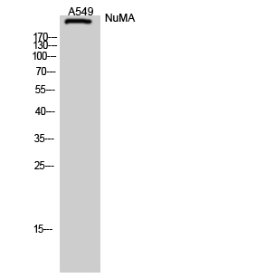
Figure 1. Western blot analysis of NUMA/NUMA1 using anti-NUMA/NUMA1 antibody (A02018-1). Electrophoresis was performed on a 5-20% SDS-PAGE gel at 70V (Stacking gel) / 90V (Resolving gel) for 2-3 hours. The sample well of each lane was loaded with 30 ug of sample under reducing conditions. Lane 1: human Hela whole cell lysates, Lane 2: monkey COS-7 whole cell lysates, Lane 3: human MCF-7 whole cell lysates, Lane 4: human SH-SY5Y whole cell lysates, Lane 5: human SIHA whole cell lysates, Lane 6: human RT4 whole cell lysates, Lane 7: rat PC-12 whole cell lysates, Lane 8: mouse NIH/3T3 whole cell lysates. After electrophoresis, proteins were transferred to a nitrocellulose membrane at 150 mA for 50-90 minutes. Blocked the membrane with 5% non-fat milk/TBS for 1.5 hour at RT. The membrane was incubated with rabbit anti-NUMA/NUMA1 antigen affinity purified polyclonal antibody (Catalog # A02018-1) at 0.5 microg/mL overnight at 4°C, then washed with TBS-0.1%Tween 3 times with 5 minutes each and probed with a goat anti-rabbit IgG-HRP secondary antibody at a dilution of 1:5000 for 1.5 hour at RT. The signal is developed using an Enhanced Chemiluminescent detection (ECL) kit (Catalog # EK1002) with Tanon 5200 system. A specific band was detected for NUMA/NUMA1 at approximately 270 kDa. The expected band size for NUMA/NUMA1 is at 270 kDa.
. NUMA/NUMA1 was detected in a paraffin-embedded section of human acinar adenocarcinoma of prostate tissue. Heat mediated antigen retrieval was performed in EDTA buffer (pH 8.0, epitope retrieval solution). The tissue section was blocked with 10% goat serum. The tissue section was then incubated with 2 microg/ml rabbit anti-NUMA/NUMA1 Antibody (A02018-1) overnight at 4°C. Peroxidase Conjugated Goat Anti-rabbit IgG was used as secondary antibody and incubated for 30 minutes at 37°C. The tissue section was developed using HRP Conjugated Rabbit IgG Super Vision Assay Kit (Catalog # SV0002) with DAB as the chromogen.")
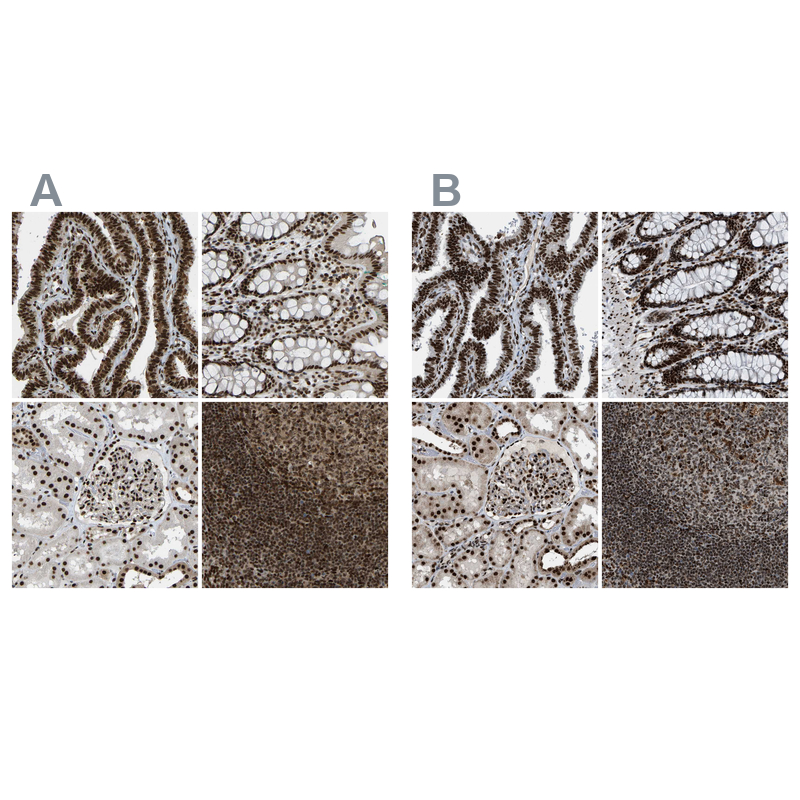
. NUMA/NUMA1 was detected in a paraffin-embedded section of human breast cancer tissue. Heat mediated antigen retrieval was performed in EDTA buffer (pH 8.0, epitope retrieval solution). The tissue section was blocked with 10% goat serum. The tissue section was then incubated with 2 microg/ml rabbit anti-NUMA/NUMA1 Antibody (A02018-1) overnight at 4°C. Peroxidase Conjugated Goat Anti-rabbit IgG was used as secondary antibody and incubated for 30 minutes at 37°C. The tissue section was developed using HRP Conjugated Rabbit IgG Super Vision Assay Kit (Catalog # SV0002) with DAB as the chromogen.")
. NUMA/NUMA1 was detected in a paraffin-embedded section of human esophageal squamous carcinoma tissue. Heat mediated antigen retrieval was performed in EDTA buffer (pH 8.0, epitope retrieval solution). The tissue section was blocked with 10% goat serum. The tissue section was then incubated with 2 microg/ml rabbit anti-NUMA/NUMA1 Antibody (A02018-1) overnight at 4°C. Peroxidase Conjugated Goat Anti-rabbit IgG was used as secondary antibody and incubated for 30 minutes at 37°C. The tissue section was developed using HRP Conjugated Rabbit IgG Super Vision Assay Kit (Catalog # SV0002) with DAB as the chromogen.")
. NUMA/NUMA1 was detected in a paraffin-embedded section of human lung cancer tissue. Heat mediated antigen retrieval was performed in EDTA buffer (pH 8.0, epitope retrieval solution). The tissue section was blocked with 10% goat serum. The tissue section was then incubated with 2 microg/ml rabbit anti-NUMA/NUMA1 Antibody (A02018-1) overnight at 4°C. Peroxidase Conjugated Goat Anti-rabbit IgG was used as secondary antibody and incubated for 30 minutes at 37°C. The tissue section was developed using HRP Conjugated Rabbit IgG Super Vision Assay Kit (Catalog # SV0002) with DAB as the chromogen.")
 and anti-Beta Tubulin antibody (M01857-3). NUMA/NUMA1 was detected in immunocytochemical section of U2OS cell. Enzyme antigen retrieval was performed using IHC enzyme antigen retrieval reagent (AR0022) for 15 mins. The cells were blocked with 10% goat serum. And then incubated with 5 microg/mL rabbit anti-NUMA/NUMA1 Antibody (A02018-1) and mouse anti-Beta Tubulin antibody (M01857-3) overnight at 4°C. Cy3 Conjugated Goat Anti-Rabbit IgG (BA1032) and DyLight®488 Conjugated Goat Anti-Mouse IgG (BA1126) were used as secondary antibody at 1:500 dilution and incubated for 30 minutes at 37°C. Visualize using a fluorescence microscope and filter sets appropriate for the label used.")
. Overlay histogram showing RT4 cells stained with A02018-1 (Blue line). To facilitate intracellular staining, cells were fixed with 4% paraformaldehyde and permeabilized with permeabilization buffer. The cells were blocked with 10% normal goat serum. And then incubated with rabbit anti-NUMA/NUMA1 Antibody (A02018-1, 1 microg/1x106 cells) for 30 min at 20°C. DyLight®488 conjugated goat anti-rabbit IgG (BA1127, 5-10 microg/1x106 cells) was used as secondary antibody for 30 minutes at 20°C. Isotype control antibody (Green line) was rabbit IgG (1 microg/1x106) used under the same conditions. Unlabelled sample (Red line) was also used as a control.")
Figure 1. Western blot analysis of NUMA/NUMA1 using anti-NUMA/NUMA1 antibody (A02018-1). Electrophoresis was performed on a 5-20% SDS-PAGE gel at 70V (Stacking gel) / 90V (Resolving gel) for 2-3 hours. The sample well of each lane was loaded with 30 ug of sample under reducing conditions. Lane 1: human Hela whole cell lysates, Lane 2: monkey COS-7 whole cell lysates, Lane 3: human MCF-7 whole cell lysates, Lane 4: human SH-SY5Y whole cell lysates, Lane 5: human SIHA whole cell lysates, Lane 6: human RT4 whole cell lysates, Lane 7: rat PC-12 whole cell lysates, Lane 8: mouse NIH/3T3 whole cell lysates. After electrophoresis, proteins were transferred to a nitrocellulose membrane at 150 mA for 50-90 minutes. Blocked the membrane with 5% non-fat milk/TBS for 1.5 hour at RT. The membrane was incubated with rabbit anti-NUMA/NUMA1 antigen affinity purified polyclonal antibody (Catalog # A02018-1) at 0.5 microg/mL overnight at 4°C, then washed with TBS-0.1%Tween 3 times with 5 minutes each and probed with a goat anti-rabbit IgG-HRP secondary antibody at a dilution of 1:5000 for 1.5 hour at RT. The signal is developed using an Enhanced Chemiluminescent detection (ECL) kit (Catalog # EK1002) with Tanon 5200 system. A specific band was detected for NUMA/NUMA1 at approximately 270 kDa. The expected band size for NUMA/NUMA1 is at 270 kDa.
Anti-NUMA/NUMA1 Antibody Picoband(r)
A02018-1-CARRIER-FREE
ApplicationsFlow Cytometry, ImmunoFluorescence, Western Blot, ELISA, ImmunoCytoChemistry, ImmunoHistoChemistry
Product group Antibodies
ReactivityHuman, Monkey, Mouse, Rat
TargetNUMA1
Overview
- SupplierBoster Bio
- Product NameAnti-NUMA/NUMA1 Antibody Picoband(r)
- Delivery Days Customer9
- ApplicationsFlow Cytometry, ImmunoFluorescence, Western Blot, ELISA, ImmunoCytoChemistry, ImmunoHistoChemistry
- CertificationResearch Use Only
- ClonalityPolyclonal
- Concentration500 ug/ml
- Gene ID4926
- Target nameNUMA1
- Target descriptionnuclear mitotic apparatus protein 1
- Target synonymsNMP-22, NUMA, nuclear mitotic apparatus protein 1, SP-H antigen, centrophilin stabilizes mitotic spindle in mitotic cells, nuclear matrix protein-22, structural nuclear protein
- HostRabbit
- IsotypeIgG
- Protein IDQ14980
- Protein NameNuclear mitotic apparatus protein 1
- Scientific DescriptionBoster Bio Anti-NUMA/NUMA1 Antibody Picoband® catalog # A02018-1. Tested in ELISA, IF, IHC, ICC, WB, Flow Cytometry applications. This antibody reacts with Human, Mouse, Rat, Monkey. The brand Picoband indicates this is a premium antibody that guarantees superior quality, high affinity, and strong signals with minimal background in Western blot applications. Only our best-performing antibodies are designated as Picoband, ensuring unmatched performance.
- ReactivityHuman, Monkey, Mouse, Rat
- Storage Instruction-20°C,2°C to 8°C
- UNSPSC12352203
Related products
Product group Antibodies
Anti-NUMA1 AntibodyA99616
ApplicationsWestern Blot, ELISA, ImmunoHistoChemistry
ReactivityHuman
- SizePrice
Product group Antibodies
Anti-NUMA1 Antibody144-00527
ApplicationsImmunoFluorescence, Western Blot, ImmunoHistoChemistry
ReactivityHuman, Mouse, Rat
TargetNUMA1
- SizePrice
Product group Antibodies
NUMA1 Recombinant Antibody, Biotin ConjugatedBSM-61733R-BIOTIN
ApplicationsWestern Blot, ELISA, ImmunoHistoChemistry, ImmunoHistoChemistry Frozen, ImmunoHistoChemistry Paraffin
ReactivityHuman, Mouse, Rat
TargetNUMA1
- SizePrice
Product group Antibodies
NUMA1 Polyclonal AntibodyCAC12913
ApplicationsImmunoFluorescence, Western Blot, ELISA, ImmunoHistoChemistry
TargetNUMA1
- SizePrice
Product group Antibodies
NUMA1 AntibodyCSB-PA016185YA01HU
ApplicationsImmunoFluorescence, Western Blot, ELISA, ImmunoHistoChemistry
ReactivityHuman
TargetNUMA1
- SizePrice
Product group Antibodies

NUMA1 / NUMA Antibody (N-Terminus)LS-C368913
ApplicationsWestern Blot, ImmunoHistoChemistry, ImmunoHistoChemistry Paraffin
ReactivityHuman
TargetNUMA1
- SizePrice
![NuMA antibody [N1], N-term detects NuMA protein at nucleus in human colon carcinoma by immunohistochemical analysis. Sample: Paraffin-embedded human colon carcinoma. NuMA antibody [N1], N-term (GTX113510) diluted at 1:500.
Antigen Retrieval: Citrate buffer, pH 6.0, 15 min](https://www.genetex.com/upload/website/prouct_img/normal/GTX113510/GTX113510_40142_20150803_IHC-P_w_23060501_408.webp)
Product group Antibodies
NuMA antibody [N1], N-termGTX113510
ApplicationsImmunoFluorescence, Western Blot, ImmunoCytoChemistry, ImmunoHistoChemistry, ImmunoHistoChemistry Paraffin
ReactivityHuman
TargetNUMA1
- SizePrice
Product group Antibodies
TargetNUMA1
- SizePrice
Product group Antibodies
Anti-NUMA1 AntibodyHPA019859
ApplicationsImmunoHistoChemistry
ReactivityHuman
TargetNUMA1
- SizePrice