

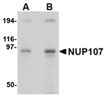

Anti-NUP107 Antibody
A48748
ApplicationsWestern Blot, ELISA, ImmunoCytoChemistry
Product group Antibodies
ReactivityHuman, Mouse, Rat
Overview
- SupplierAntibodies.com
- Product NameAnti-NUP107 Antibody
- Delivery Days Customer7
- ApplicationsWestern Blot, ELISA, ImmunoCytoChemistry
- CertificationResearch Use Only
- ClonalityPolyclonal
- ConjugateUnconjugated
- HostRabbit
- Scientific DescriptionRabbit polyclonal antibody to NUP107
- ReactivityHuman, Mouse, Rat
- UNSPSC12352203
Related products
Product group Antibodies
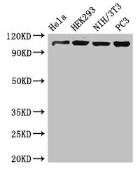
NUP107 AntibodyCSB-PA016188LA01HU
ApplicationsWestern Blot, ELISA, ImmunoHistoChemistry
ReactivityHuman, Mouse
TargetNUP107
- SizePrice
Product group Antibodies

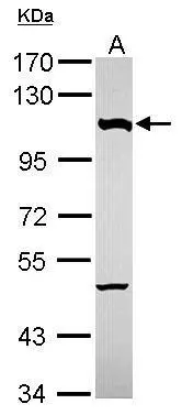
NUP107 AntibodyLS-C748170
ApplicationsWestern Blot
ReactivityHuman, Mouse
TargetNUP107
- SizePrice
Product group Antibodies

Goat anti-NUP107EB10160
ApplicationsWestern Blot, ELISA
ReactivityBovine, Canine, Human, Mouse, Rat
TargetNUP107
- SizePrice

Product group Antibodies
References
Anti-NUP107 Antibody Picoband(r)A03724-1-CARRIER-FREE
ApplicationsFlow Cytometry, ImmunoFluorescence, ImmunoPrecipitation, Western Blot, ELISA, ImmunoCytoChemistry
ReactivityHuman, Mouse, Rat
TargetNUP107
- SizePrice
Product group Antibodies
NUP107 Polyclonal AntibodyCAC12914
ApplicationsWestern Blot, ELISA, ImmunoHistoChemistry
ReactivityMouse
TargetNUP107
- SizePrice
Product group Antibodies
NUP107 antibodyGTX116664
ApplicationsImmunoPrecipitation, Western Blot
ReactivityHuman
TargetNUP107
- SizePrice
Product group Antibodies
Anti-NUP107 Antibody144-60193
ApplicationsWestern Blot
ReactivityHuman, Mouse
TargetNUP107
- SizePrice
Product group Antibodies
NUP107 Recombinant AntibodyBSM-62690R
ApplicationsWestern Blot
ReactivityHuman
TargetNUP107
- SizePrice