

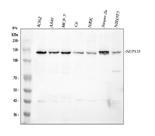
Figure 1. Western blot analysis of NUP133 using anti-NUP133 antibody (A05327-2). Electrophoresis was performed on a 5-20% SDS-PAGE gel at 70V (Stacking gel) / 90V (Resolving gel) for 2-3 hours. The sample well of each lane was loaded with 30 ug of sample under reducing conditions. Lane 1: human K562 whole cell lysates, Lane 2: human A549 whole cell lysates, Lane 3: human MCF-7 whole cell lysates, Lane 4: rat C6 whole cell lysates, Lane 5: rat NRK whole cell lysates, Lane 6: mouse Neuro-2a whole cell lysates, Lane 7: mouse NIH/3T3 whole cell lysates. After electrophoresis, proteins were transferred to a nitrocellulose membrane at 150 mA for 50-90 minutes. Blocked the membrane with 5% non-fat milk/TBS for 1.5 hour at RT. The membrane was incubated with rabbit anti-NUP133 antigen affinity purified polyclonal antibody (Catalog # A05327-2) at 0.5 microg/mL overnight at 4°C, then washed with TBS-0.1%Tween 3 times with 5 minutes each and probed with a goat anti-rabbit IgG-HRP secondary antibody at a dilution of 1:5000 for 1.5 hour at RT. The signal is developed using an Enhanced Chemiluminescent detection (ECL) kit (Catalog # EK1002) with Tanon 5200 system. A specific band was detected for NUP133 at approximately 129 kDa. The expected band size for NUP133 is at 129 kDa.
 and anti-Beta Tubulin antibody (M01857-3). NUP133 was detected in immunocytochemical section of A549 cell. Enzyme antigen retrieval was performed using IHC enzyme antigen retrieval reagent (AR0022) for 15 mins. The cells were blocked with 10% goat serum. And then incubated with 5 microg/mL rabbit anti-NUP133 Antibody (A05327-2) and mouse anti-Beta Tubulin antibody (M01857-3) overnight at 4°C. DyLight®488 Conjugated Goat Anti-Rabbit IgG (BA1127) and Cy3 Conjugated Conjugated Goat Anti-Mouse IgG (BA1031) were used as secondary antibody at 1:500 dilution and incubated for 30 minutes at 37°C. Visualize using a fluorescence microscope and filter sets appropriate for the label used.")
 and anti-Beta Tubulin antibody (M01857-3). NUP133 was detected in immunocytochemical section of C6 cell. Enzyme antigen retrieval was performed using IHC enzyme antigen retrieval reagent (AR0022) for 15 mins. The cells were blocked with 10% goat serum. And then incubated with 5 microg/mL rabbit anti-NUP133 Antibody (A05327-2) and mouse anti-Beta Tubulin antibody (M01857-3) overnight at 4°C. Cy3 Conjugated Goat Anti-Rabbit IgG (BA1032) and DyLight®488 Conjugated Conjugated Goat Anti-Mouse IgG (BA1126) were used as secondary antibody at 1:500 dilution and incubated for 30 minutes at 37°C. Visualize using a fluorescence microscope and filter sets appropriate for the label used.")
. Lane 1: Hela whole cell lysates (30ug) Lane 2: Rabbit control IgG instead of anti-NUP133 antibody in Hela whole cell lysate. Lane 3: anti-NUP133 antibody (2microg) + Hela whole cell lysate (500microg) After electrophoresis, proteins were transferred to a membrane. Then the membrane was incubated with rabbit anti-NUP133 antigen affinity purified polyclonal antibody (A05327-2) at a dilution of 0.5 microg/mL and probed with a goat anti-rabbit IgG-HRP secondary antibody (Heavy Chain). The signal is developed using ECL Plus Western Blotting Substrate (Catalog # AR1197). A specific band was detected for NUP133 at approximately 129 kDa. The expected band size for NUP133 is at 129 kDa.")
Figure 1. Western blot analysis of NUP133 using anti-NUP133 antibody (A05327-2). Electrophoresis was performed on a 5-20% SDS-PAGE gel at 70V (Stacking gel) / 90V (Resolving gel) for 2-3 hours. The sample well of each lane was loaded with 30 ug of sample under reducing conditions. Lane 1: human K562 whole cell lysates, Lane 2: human A549 whole cell lysates, Lane 3: human MCF-7 whole cell lysates, Lane 4: rat C6 whole cell lysates, Lane 5: rat NRK whole cell lysates, Lane 6: mouse Neuro-2a whole cell lysates, Lane 7: mouse NIH/3T3 whole cell lysates. After electrophoresis, proteins were transferred to a nitrocellulose membrane at 150 mA for 50-90 minutes. Blocked the membrane with 5% non-fat milk/TBS for 1.5 hour at RT. The membrane was incubated with rabbit anti-NUP133 antigen affinity purified polyclonal antibody (Catalog # A05327-2) at 0.5 microg/mL overnight at 4°C, then washed with TBS-0.1%Tween 3 times with 5 minutes each and probed with a goat anti-rabbit IgG-HRP secondary antibody at a dilution of 1:5000 for 1.5 hour at RT. The signal is developed using an Enhanced Chemiluminescent detection (ECL) kit (Catalog # EK1002) with Tanon 5200 system. A specific band was detected for NUP133 at approximately 129 kDa. The expected band size for NUP133 is at 129 kDa.
Anti-NUP133 Antibody Picoband(r)
A05327-2-CARRIER-FREE
ApplicationsImmunoFluorescence, ImmunoPrecipitation, Western Blot, ELISA, ImmunoCytoChemistry
Product group Antibodies
ReactivityHuman, Mouse, Rat
TargetNUP133
Overview
- SupplierBoster Bio
- Product NameAnti-NUP133 Antibody Picoband(r)
- Delivery Days Customer9
- ApplicationsImmunoFluorescence, ImmunoPrecipitation, Western Blot, ELISA, ImmunoCytoChemistry
- CertificationResearch Use Only
- ClonalityPolyclonal
- Concentration500 ug/ml
- Gene ID55746
- Target nameNUP133
- Target descriptionnucleoporin 133
- Target synonymsGAMOS8, NPHS18, hNUP133, nuclear pore complex protein Nup133, 133 kDa nucleoporin, nucleoporin 133kD, nucleoporin 133kDa, nucleoporin Nup133
- HostRabbit
- IsotypeIgG
- Protein IDQ8WUM0
- Protein NameNuclear pore complex protein Nup133
- Scientific DescriptionBoster Bio Anti-NUP133 Antibody Picoband® catalog # A05327-2. Tested in ELISA, IP, IF, ICC, WB applications. This antibody reacts with Human, Mouse, Rat. The brand Picoband indicates this is a premium antibody that guarantees superior quality, high affinity, and strong signals with minimal background in Western blot applications. Only our best-performing antibodies are designated as Picoband, ensuring unmatched performance.
- ReactivityHuman, Mouse, Rat
- Storage Instruction-20°C,2°C to 8°C
- UNSPSC12352203
Related products
Product group Antibodies
NUP133 AntibodyCSB-PA016189GA01HU
ApplicationsWestern Blot, ELISA, ImmunoHistoChemistry
ReactivityHuman, Mouse, Rat
TargetNUP133
- SizePrice
Product group Antibodies
Anti-NUP133 AntibodyA10522
ApplicationsImmunoFluorescence, Western Blot, ImmunoCytoChemistry
ReactivityHuman
- SizePrice

Product group Antibodies
Anti-NUP133-25ulHPA059767
ApplicationsWestern Blot, ImmunoCytoChemistry, ImmunoHistoChemistry
ReactivityHuman
- SizePrice
Product group Antibodies

NUP133 AntibodyLS-C410352
ApplicationsWestern Blot
ReactivityHuman
TargetNUP133
- SizePrice
Product group Antibodies
ApplicationsImmunoPrecipitation, Western Blot, ImmunoCytoChemistry, ImmunoHistoChemistry
TargetNUP133
- SizePrice

Product group Antibodies
NUP133 antibodyGTX64772
ApplicationsWestern Blot
ReactivityHuman
TargetNUP133
- SizePrice
Product group Antibodies
Anti-NUP133 Antibody144-08818
ApplicationsWestern Blot
ReactivityHuman
TargetNUP133
- SizePrice