

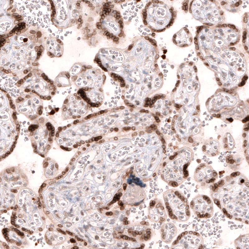
Immunohistochemical staining of human placenta shows moderate to strong nuclear membrane positivity in trophoblastic cells.

Immunohistochemical staining of human placenta shows moderate to strong nuclear membrane positivity in trophoblastic cells.
Anti-NUP54-25ul
HPA035929
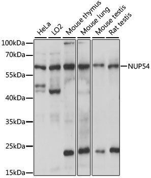
ApplicationsWestern Blot, ImmunoHistoChemistry
Product group Antibodies
ReactivityHuman, Mouse, Rat
Overview
- SupplierAtlas Antibodies
- Product NameAnti-NUP54-25ul
- Delivery Days Customer6
- ApplicationsWestern Blot, ImmunoHistoChemistry
- Applications SupplierIHC, WB
- CertificationResearch Use Only
- ClonalityPolyclonal
- Concentration0.1
- ConjugateUnconjugated
- HostRabbit
- IsotypeIgG
- Protein IDQ7Z3B4
- Protein NameNucleoporin p54
- Scientific DescriptionRabbit Polyclonal Anti-NUP54 Antibody against Human nucleoporin 54kDa. Validated for Immunohistochemistry and Western Blot
- ReactivityHuman, Mouse, Rat
- Storage InstructionStore at +4°C for short term storage. Long time storage is recommended at -20°C.
- UNSPSC12352203
Datasheet
MSDS
Related products
Product group Antibodies
Anti-NUP54 Antibody144-61064
ApplicationsWestern Blot
ReactivityHuman, Mouse, Rat
TargetNUP54
- SizePrice
Product group Antibodies
NUP54 AntibodyCSB-PA016203GA01HU
ApplicationsWestern Blot, ELISA
ReactivityHuman, Mouse, Rat
TargetNUP54
- SizePrice
Product group Antibodies

NUP54 AntibodyLS-C750424
ApplicationsWestern Blot
ReactivityHuman, Mouse, Rat
TargetNUP54
- SizePrice
Product group Antibodies
Anti-NUP54 AntibodyA90657
ApplicationsWestern Blot
ReactivityHuman, Mouse, Rat
- SizePrice
Product group Antibodies
NUP54 Polyclonal AntibodyBS-4028R
ApplicationsImmunoFluorescence, ELISA, ImmunoCytoChemistry, ImmunoHistoChemistry, ImmunoHistoChemistry Frozen, ImmunoHistoChemistry Paraffin
ReactivityBovine, Chicken, Equine, Human, Mouse, Porcine, Rabbit, Rat
TargetNUP54
- SizePrice

Product group Antibodies
Anti-NUP54 AntibodyHPA058122
ApplicationsImmunoCytoChemistry
ReactivityHuman
TargetNUP54
- SizePrice
Product group Antibodies
Anti-NUP54 AntibodyHPA058122
ApplicationsImmunoCytoChemistry
ReactivityHuman
TargetNUP54
- SizePrice

Product group Antibodies
Anti-NUP54 Antibody Picoband(r)A10830-1-CARRIER-FREE
ApplicationsWestern Blot, ELISA
ReactivityHuman
TargetNUP54
- SizePrice