

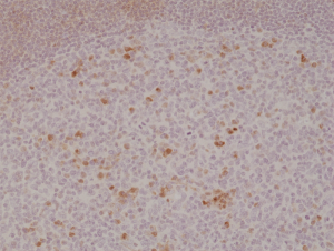
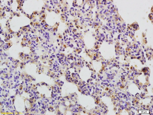
Immunohistochemical staining of formalin fixed and paraffin embedded human tonsil tissue section using anti-OX40 rabbit monoclonal antibody (Clone RM313) at a 1:500 dilution.
Immunohistochemical staining of formalin fixed and paraffin embedded human tonsil tissue section using anti-OX40 rabbit monoclonal antibody (Clone RM313) at a 1:500 dilution.
anti-OX40 (human), Rabbit Monoclonal (RM313)
REV-31-1199-00
ApplicationsWestern Blot, ImmunoHistoChemistry
Product group Antibodies
ReactivityHuman
TargetTNFRSF4
Overview
- SupplierRevMAb Biosciences
- Product Nameanti-OX40 (human), Rabbit Monoclonal (RM313)
- Delivery Days Customer10
- ApplicationsWestern Blot, ImmunoHistoChemistry
- CertificationResearch Use Only
- ClonalityMonoclonal
- Clone IDRM313
- Gene ID7293
- Target nameTNFRSF4
- Target descriptionTNF receptor superfamily member 4
- Target synonymsACT35, CD134, IMD16, OX40, TXGP1L, tumor necrosis factor receptor superfamily member 4, ACT35 antigen, ATC35 antigen, CD134 antigen, OX40 antigen, OX40 cell surface antigen, OX40 homologue, OX40L receptor, TAX transcriptionally-activated glycoprotein 1 receptor, lymphoid activation antigene ACT35, tax-transcriptionally activated glycoprotein 1 receptor
- HostRabbit
- IsotypeIgG
- Protein IDP43489
- Protein NameTumor necrosis factor receptor superfamily member 4
- Scientific DescriptionOX40 receptor is a member of the TNFR-superfamily of receptors which is not constitutively expressed on resting naive T cells. OX40 is a secondary co-stimulatory immune checkpoint molecule. OX40 binds to receptors on T-cells, preventing them from dying and subsequently increasing cytokine production. OX40 has a critical role in the maintenance of an immune response. OX40 expression can promote viral binding and renders cells permissive for viral entry, productive infection and syncytium formation. Stimulating the receptor can improve the response to a powerful virus vector and may be useful in vaccine development. - Recombinant Antibody. This antibody reacts to human OX40 (CD134). Applications: WB, IHC. Source: Rabbit. Liquid. 50% Glycerol/PBS with 1% BSA and 0.09% sodium azide. OX40 receptor is a member of the TNFR-superfamily of receptors which is not constitutively expressed on resting naive T cells. OX40 is a secondary co-stimulatory immune checkpoint molecule. OX40 binds to receptors on T-cells, preventing them from dying and subsequently increasing cytokine production. OX40 has a critical role in the maintenance of an immune response. OX40 expression can promote viral binding and renders cells permissive for viral entry, productive infection and syncytium formation. Stimulating the receptor can improve the response to a powerful virus vector and may be useful in vaccine development.
- ReactivityHuman
- Storage Instruction-20°C,2°C to 8°C
- UNSPSC41116161
Datasheet
Related products
Product group Antibodies

Anti-CD134 [A10-F]Ab02228-10.0
ApplicationsFlow Cytometry, ELISA, Neutralisation/Blocking
ReactivityHuman, Monkey, Primate
TargetTNFRSF4
- SizePrice
Product group Antibodies

OX40 AntibodyABX037462
ApplicationsWestern Blot, ELISA, ImmunoHistoChemistry
- SizePrice
Product group Antibodies

ApplicationsFlow Cytometry
ReactivityHuman
TargetTNFRSF4
- SizePrice
Product group Antibodies
CD134 Recombinant AntibodyBSM-60298R
ApplicationsFlow Cytometry, ImmunoFluorescence, Western Blot, ImmunoCytoChemistry, ImmunoHistoChemistry, ImmunoHistoChemistry Frozen, ImmunoHistoChemistry Paraffin
ReactivityHuman, Mouse, Rat
TargetTNFRSF4
- SizePrice
Product group Antibodies
TNFRSF4 AntibodyCSB-PA006263
ApplicationsWestern Blot, ELISA
ReactivityHuman
TargetTNFRSF4
- SizePrice
Product group Antibodies
ApplicationsELISA, ImmunoHistoChemistry
TargetTNFRSF4
- SizePrice
Product group Antibodies
CD134 antibodyGTX51548
ApplicationsImmunoHistoChemistry, ImmunoHistoChemistry Paraffin
ReactivityHuman, Rat
TargetTNFRSF4
- SizePrice
Product group Antibodies
TargetTNFRSF4
- SizePrice

Product group Antibodies
Anti-CD134/OX40/TNFRSF4 Antibody Picoband(r)PB9898-CARRIER-FREE
ApplicationsFlow Cytometry, Western Blot, ImmunoCytoChemistry, ImmunoHistoChemistry
ReactivityHuman
TargetTNFRSF4
- SizePrice