

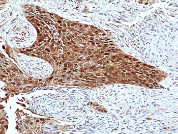
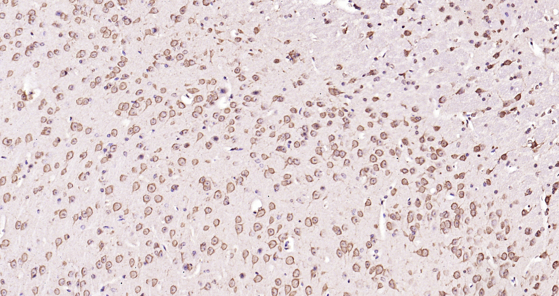
Immunohistochemical staining of formalin-fixed and paraffin-embedded human Lung Squamous Carcinoma tissue sections using Anti-Erk2 antibody (RM483) at 1:100 dilution.
Immunohistochemical staining of formalin-fixed and paraffin-embedded human Lung Squamous Carcinoma tissue sections using Anti-Erk2 antibody (RM483) at 1:100 dilution.
anti-p42 MAPK (Erk2), Rabbit Monoclonal (RM483)
REV-31-1375-00
ApplicationsWestern Blot, ImmunoHistoChemistry
Product group Antibodies
ReactivityHuman, Mouse, Rat
TargetMAPK1
Overview
- SupplierRevMAb Biosciences
- Product Nameanti-p42 MAPK (Erk2), Rabbit Monoclonal (RM483)
- Delivery Days Customer10
- ApplicationsWestern Blot, ImmunoHistoChemistry
- CertificationResearch Use Only
- ClonalityMonoclonal
- Clone IDRM483
- Gene ID5594
- Target nameMAPK1
- Target descriptionmitogen-activated protein kinase 1
- Target synonymsERK, ERK-2, ERK2, ERT1, MAPK2, NS13, P42MAPK, PRKM1, PRKM2, p38, p40, p41, p41mapk, p42-MAPK, mitogen-activated protein kinase 1, MAP kinase 1, MAP kinase 2, MAPK 2, extracellular signal-regulated kinase 2, mitogen-activated protein kinase 2, protein tyrosine kinase ERK2
- HostRabbit
- IsotypeIgG
- Protein IDP28482
- Protein NameMitogen-activated protein kinase 1
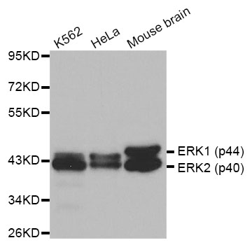
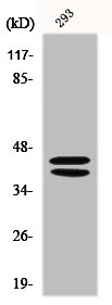
- Scientific DescriptionExtracellular signal-regulated kinases 1 and 2 (also known as ERK1/2, p44/42, or MAPK3/1) belong to a family of conserved serine/threonine protein kinases known as mitogen-activated protein kinases (MAPKs) that are involved in many cellular programs such as proliferation, differentiation, motility and survival. The ERK1/2 signaling pathway can be activated in response to a diverse range of extracellular stimuli including mitogens, growth factors and cytokines. The primary activators of ERK1/2 are MEK1 and MEK2 which activate p44 and p42 through phosphorylation of activation loop residues Thr202/Tyr204 and Thr185/Tyr187 in ERK1 and ERK2, respectively. Several downstream targets of ERK1/2 have been identified, including p90RSK and the transcription factor Elk-1. ERK1/2 are negatively regulated by MAPK phosphatases as well as by chemical inhibitors of MEK. The ERK pathway is disrupted in many types of cancer and thus is an important target for diagnosis and treatment. - Recombinant Antibody. This antibody reacts to human p42 MAPK (Erk2). This antibody may also react to mouse or rat Erk2 as predicted by immunogen homology. Source: Rabbit. Isotype: Rabbit IgG. Immunogen: A peptide corresponding to the C-terminus of human p42-MAPK (Erk2). Applications: IHC, WB. Extracellular signal-regulated kinases 1 and 2 (also known as ERK1/2, p44/42, or MAPK3/1) belong to a family of conserved serine/threonine protein kinases known as mitogen-activated protein kinases (MAPKs) that are involved in many cellular programs such as proliferation, differentiation, motility and survival. The ERK1/2 signaling pathway can be activated in response to a diverse range of extracellular stimuli including mitogens, growth factors and cytokines. The primary activators of ERK1/2 are MEK1 and MEK2 which activate p44 and p42 through phosphorylation of activation loop residues Thr202/Tyr204 and Thr185/Tyr187 in ERK1 and ERK2, respectively. Several downstream targets of ERK1/2 have been identified, including p90RSK and the transcription factor Elk-1. ERK1/2 are negatively regulated by MAPK phosphatases as well as by chemical inhibitors of MEK. The ERK pathway is disrupted in many types of cancer and thus is an important target for diagnosis and treatment.
- ReactivityHuman, Mouse, Rat
- Storage Instruction-20°C,2°C to 8°C
- UNSPSC41116161
Datasheet
Related products
Product group Antibodies
Anti-ERK1/2 AntibodyA29693
ApplicationsImmunoFluorescence, Western Blot, ImmunoHistoChemistry
ReactivityHuman, Mouse, Rat
- SizePrice
Product group Antibodies
Anti-ERK1 / ERK2 Antibody144-64587
ApplicationsImmunoFluorescence, Western Blot, ImmunoHistoChemistry
ReactivityHuman, Mouse, Rat
TargetMAPK1
- SizePrice

Product group Antibodies
References
ApplicationsFlow Cytometry, ImmunoFluorescence, ImmunoPrecipitation, Western Blot, ImmunoCytoChemistry, ImmunoHistoChemistry
ReactivityHuman, Mouse, Rat
TargetMAPK1
- SizePrice
Product group Antibodies
MAPK3/MAPK1 AntibodyCSB-PA002419
ApplicationsImmunoFluorescence, Western Blot, ELISA
ReactivityHuman, Mouse, Rat
TargetMAPK1
- SizePrice
Product group Antibodies

Goat anti-ERK2 / MAPK1EB06592
ApplicationsImmunoFluorescence, Western Blot, ELISA
ReactivityBovine, Canine, Human, Mouse, Rat
TargetMAPK1
- SizePrice
Product group Antibodies
MAPK1 Polyclonal AntibodyCAC14542
ApplicationsImmunoFluorescence, Western Blot, ELISA, ImmunoHistoChemistry
ReactivityRat
TargetMAPK1
- SizePrice
Product group Antibodies
References
ApplicationsFlow Cytometry, ImmunoFluorescence, Western Blot, ELISA, ImmunoCytoChemistry, ImmunoHistoChemistry, ImmunoHistoChemistry Frozen, ImmunoHistoChemistry Paraffin
ReactivityBovine, Canine, Chicken, Equine, Guinea Pig, Human, Mouse, Porcine, Rabbit, Rat
TargetMAPK1
- SizePrice
Product group Antibodies

ApplicationsELISA
ReactivityHuman
TargetMAPK1
- SizePrice

Product group Antibodies
Anti-MAPK1-25ulHPA030069
ApplicationsWestern Blot, ImmunoHistoChemistry
ReactivityHuman
- SizePrice