


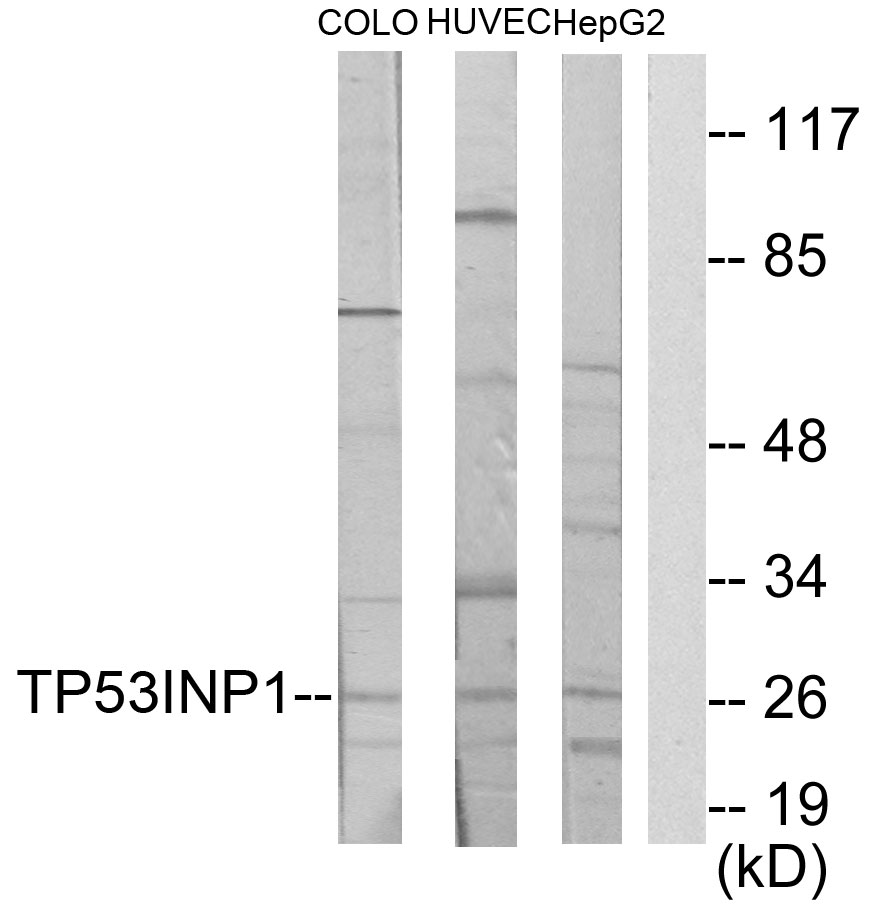
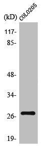
Western blot analysis of p53 DINP1 expression in (1) HepG2 cell lysate; (2) RAW 264.7 cell lysate; (3) PC12 cell lysate.
Western blot analysis of p53 DINP1 expression in (1) HepG2 cell lysate; (2) RAW 264.7 cell lysate; (3) PC12 cell lysate.
Anti-p53 DINP1 Rabbit Monoclonal Antibody
M04229-1
ApplicationsImmunoFluorescence, Western Blot, ImmunoCytoChemistry, ImmunoHistoChemistry
Product group Antibodies
ReactivityHuman, Mouse, Rat
TargetTP53INP1
Overview
- SupplierBoster Bio
- Product NameAnti-p53 DINP1 Rabbit Monoclonal Antibody
- Delivery Days Customer9
- ApplicationsImmunoFluorescence, Western Blot, ImmunoCytoChemistry, ImmunoHistoChemistry
- CertificationResearch Use Only
- ClonalityMonoclonal
- Clone ID19T28
- Gene ID94241
- Target nameTP53INP1
- Target descriptiontumor protein p53 inducible nuclear protein 1
- Target synonymsSIP, TP53DINP1, TP53INP1A, TP53INP1B, Teap, p53DINP1, tumor protein p53-inducible nuclear protein 1, p53-dependent damage-inducible nuclear protein 1, p53-inducible p53DINP1, stress-induced protein
- HostRabbit
- IsotypeIgG
- Protein IDQ96A56
- Protein NameTumor protein p53-inducible nuclear protein 1
- Scientific DescriptionBoster Bio Anti-p53 DINP1 Rabbit Monoclonal Antibody catalog # M04229-1. Tested in WB, IHC, ICC/IF applications. This antibody reacts with Human, Mouse, Rat.
- ReactivityHuman, Mouse, Rat
- Storage Instruction-20°C
- UNSPSC12352203
References
- Fu R, Dou Z, Li N, et al. Avenanthramide C induces cellular senescence in colorectal cancer cells via suppressing β-catenin-mediated the transcription of miR-183/96/182 cluster. Biochem Pharmacol. 2022,199:115021. doi: 10.1016/j.bcp.2022.115021Read this paper
- Li Y, Liu M, Song X, et al. Exogenous Hydrogen Sulfide Ameliorates Diabetic Myocardial Fibrosis by Inhibiting Cell Aging Through SIRT6/AMPK Autophagy. Front Pharmacol. 2020,11:1150. doi: 10.3389/fphar.2020.01150Read this paper
- Zhan X , Guan YQ. Design of magnetic nanoparticles for hepatocellular carcinoma treatment using the control mechanisms of the cell internal nucleus and external membrane. J Mater Chem B. 2015,3(20):4191-4204. doi: 10.1039/c5tb00514kRead this paper
- Li X, Sun D, Zhao T, et al. Long non-coding RNA ROR confers arsenic trioxide resistance to HepG2 cells by inhibiting p53 expression. Eur J Pharmacol. 2020,872:172982. doi: 10.1016/j.ejphar.2020.172982Read this paper
- Yabasin IB, Sanches JGP, Ibrahim MM, et al. Cisatracurium Retards Cell Migration and Invasion Upon Upregulation of p53 and Inhibits the Aggressiveness of Colorectal Cancer. Front Physiol. 2018,9:941. doi: 10.3389/fphys.2018.00941Read this paper
- Song ZH, Wang HM, Liu M, et al. Involvement of S100A4/Mts1 and associated proteins in the protective effect of fluoxetine against MCT - Induced pulmonary hypertension in rats. J Chin Med Assoc. 2018,81(12):1077-1087. doi: 10.1016/j.jcma.2018.03.013Read this paper
- Wang J, Dong B, Yu ZX, et al. The impact of acute thermal stress on green mussel Perna viridis: Oxidative damage and responses. Comp Biochem Physiol A Mol Integr Physiol. 2018,222:7-15. doi: 10.1016/j.cbpa.2018.04.001Read this paper
- Yang J, Dai LX, Chen M, et al. Inhibition of antiviral drug cidofovir on proliferation of human papillomavirus-infected cervical cancer cells. Exp Ther Med. 2016,12(5):2965-2973.Read this paper
- Xiao S, Zhou Y, Yi W, et al. Fra-1 is downregulated in cervical cancer tissues and promotes cervical cancer cell apoptosis by p53 signaling pathway in vitro. Int J Oncol. 2015,46(4):1677-84. doi: 10.3892/ijo.2015.2873Read this paper
- Gao W, Su X, Dong X, et al. Cycloartan-24-ene-1α,2α,3β-triol, a cycloartane-type triterpenoid from the resinous exudates of Commiphora myrrha, induces apoptosis in human prostatic cancer PC-3 cells. Oncol Rep. 2015,33(3):1107-14. doi: 10.3892/or.2015.3725Read this paper
Related products
Product group Antibodies
Anti-TP53INP1 AntibodyA99937
ApplicationsWestern Blot, ELISA
ReactivityHuman
- SizePrice
Product group Antibodies
Anti-TP53INP1 Antibody102-26150
ApplicationsWestern Blot
TargetTP53INP1
- SizePrice
Product group Antibodies
p53 DINP1 Recombinant Antibody, AbBy Fluor-555 ConjugatedBSM-61980R-BF555
ApplicationsImmunoFluorescence, Western Blot
ReactivityHuman, Mouse, Rat
TargetTP53INP1
- SizePrice
Product group Antibodies
TP53INP1 AntibodyCSB-PA004318
ApplicationsWestern Blot, ELISA, ImmunoHistoChemistry
ReactivityHuman
TargetTP53INP1
- SizePrice
Product group Antibodies

SIP / TP53INP1 AntibodyLS-C409809
ApplicationsWestern Blot
ReactivityHuman, Mouse, Rat
TargetTP53INP1
- SizePrice

Product group Antibodies
Anti-TP53INP1 AntibodyHPA005856
ApplicationsImmunoCytoChemistry, ImmunoHistoChemistry
ReactivityHuman
TargetTP53INP1
- SizePrice
Product group Antibodies
p53DINP1 antibody [N1N2], N-termGTX112066
ApplicationsWestern Blot, ImmunoHistoChemistry, ImmunoHistoChemistry Paraffin
ReactivityHuman
TargetTP53INP1
- SizePrice
Product group Antibodies
Anti-TP53INP1Y058708
ApplicationsWestern Blot, ELISA, ImmunoHistoChemistry
ReactivityHuman, Mouse, Rat
- SizePrice