

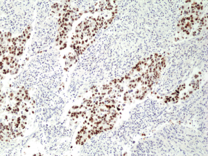
Immunohistochemical staining of formalin fixed and paraffin embedded human lung cancer tissue section using anti-p53 rabbit monoclonal antibody (Clone RM 387) at a 1:100 dilution.
Immunohistochemical staining of formalin fixed and paraffin embedded human lung cancer tissue section using anti-p53 rabbit monoclonal antibody (Clone RM 387) at a 1:100 dilution.
anti-p53 (human), Rabbit Monoclonal (RM387)
REV-31-1273-00
ApplicationsWestern Blot, ImmunoHistoChemistry
Product group Antibodies
ReactivityHuman
TargetTP53
Overview
- SupplierRevMAb Biosciences
- Product Nameanti-p53 (human), Rabbit Monoclonal (RM387)
- Delivery Days Customer10
- ApplicationsWestern Blot, ImmunoHistoChemistry
- CertificationResearch Use Only
- ClonalityMonoclonal
- Clone IDRM387
- Gene ID7157
- Target nameTP53
- Target descriptiontumor protein p53
- Target synonymsBCC7, BMFS5, LFS1, P53, TRP53, cellular tumor antigen p53, antigen NY-CO-13, mutant tumor protein 53, phosphoprotein p53, transformation-related protein 53, tumor protein 53, tumor supressor p53
- HostRabbit
- IsotypeIgG
- Protein IDP04637
- Protein NameCellular tumor antigen p53
- Scientific DescriptionRecombinant Antibody. This antibody reacts to human p53. Applications: WB, IHC. Source: Rabbit. Liquid. 50% Glycerol/PBS with 1% BSA and 0.09% sodium azide. The tumor suppressor protein, p53, is a sequence specific transcription factor that is activated by cellular stress. p53 mediates cell cycle arrest or apoptosis in response to DNA damage or starvation for pyrimidine nucleotides. p53 is up-regulated in response to stress signals and stimulated to activate transcription of specific genes, resulting in expression of p21waf1 and other proteins involved in G1 or G2/M arrest. The structure of p53 comprises an N-terminal transactivation domain, a central DNA-binding domain, an oligomerization domain, and a C-terminal regulatory domain. There are various phosphorylation sites on p53, of which the phosphorylation at Ser15 is important for p53 activation and stabilization. p53 has been characterized to play a role in blocking the proliferative action of damaged cells and act as an anticancer agent. Phosphorylation of Ser392 in p53 has been shown to associate with the formation of human tumors. In addition, p53 has also been linked to the effects of aging and oxidative stress and an increase in p53 has been linked to deficits in LTP (Long Term Potentiation) in learning and memory. Very rare normal cells express p53, but alterations in the p53 suppressor gene result in an overproduction of this protein in malignancies. Mutants of p53 that frequently occur in a number of different human cancers fail to bind the consensus DNA binding site, and cause the loss of tumor suppressor activity. - The tumor suppressor protein, p53, is a sequence specific transcription factor that is activated by cellular stress. p53 mediates cell cycle arrest or apoptosis in response to DNA damage or starvation for pyrimidine nucleotides. p53 is up-regulated in response to stress signals and stimulated to activate transcription of specific genes, resulting in expression of p21waf1 and other proteins involved in G1 or G2/M arrest. The structure of p53 comprises an N-terminal transactivation domain, a central DNA-binding domain, an oligomerization domain, and a C-terminal regulatory domain. There are various phosphorylation sites on p53, of which the phosphorylation at Ser15 is important for p53 activation and stabilization. p53 has been characterized to play a role in blocking the proliferative action of damaged cells and act as an anticancer agent. Phosphorylation of Ser392 in p53 has been shown to associate with the formation of human tumors. In addition, p53 has also been linked to the effects of aging and oxidative stress and an increase in p53 has been linked to deficits in LTP (Long Term Potentiation) in learning and memory. Very rare normal cells express p53, but alterations in the p53 suppressor gene result in an overproduction of this protein in malignancies. Mutants of p53 that frequently occur in a number of different human cancers fail to bind the consensus DNA binding site, and cause the loss of tumor suppressor activity.
- ReactivityHuman
- Storage Instruction-20°C,2°C to 8°C
- UNSPSC41116161
Datasheet
Related products
Product group Antibodies

Anti-p53 [PAb421]Ab00142-2.0
ApplicationsImmunoFluorescence, ImmunoPrecipitation, Western Blot, ELISA, ImmunoHistoChemistry, ImmunoHistoChemistry Frozen, ImmunoHistoChemistry Paraffin, Other Application
ReactivityHuman, Monkey, Mouse, Rabbit, Rat
TargetTP53
- SizePrice
Product group Antibodies
Anti-p53 AntibodyA101571
ApplicationsELISA, ImmunoHistoChemistry
ReactivityHuman
- SizePrice

Product group Antibodies
Anti-p53 AntibodyAMAB90956
ApplicationsWestern Blot, ImmunoCytoChemistry, ImmunoHistoChemistry
ReactivityHuman
TargetTP53
- SizePrice
Product group Antibodies

ApplicationsELISA
ReactivityHuman
TargetTP53
- SizePrice

Product group Antibodies
Anti-TP53 Antibody Picoband(r)A00001-2-CARRIER-FREE
ApplicationsFlow Cytometry, Western Blot, ELISA, ImmunoHistoChemistry
ReactivityHuman
TargetTP53
- SizePrice
Product group Antibodies
References
P53 Polyclonal AntibodyBS-0033R
ApplicationsFlow Cytometry, ImmunoFluorescence, Western Blot, ELISA, ImmunoCytoChemistry, ImmunoHistoChemistry, ImmunoHistoChemistry Frozen, ImmunoHistoChemistry Paraffin
ReactivityBovine, Equine, Human, Mouse, Porcine, Rabbit, Rat, Sheep
TargetTP53
- SizePrice
Product group Antibodies
TP53 Monoclonal AntibodyCSB-MA000208
ApplicationsImmunoFluorescence, Western Blot, ELISA, ImmunoHistoChemistry
ReactivityHuman
TargetTP53
- SizePrice
Product group Antibodies
Tp53 Polyclonal AntibodyCAC07006
ApplicationsWestern Blot, ChIP Chromatin ImmunoPrecipitation, ELISA, ImmunoHistoChemistry
ReactivityMouse, Rat
TargetTP53
- SizePrice