


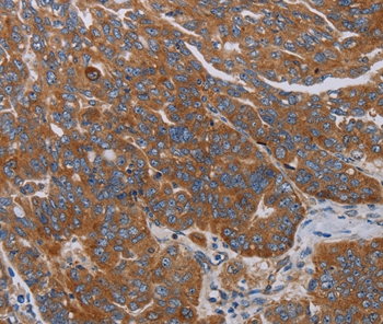
Immunohistochemical staining of human lymph node shows cytoplasmic positivity in non-germinal center cells.
![Lane 1: Marker [kDa] 230, 130, 95, 72, 56, 36, 28, 17, 11. Lane 2: Human cell line RT-4. Lane 3: Human cell line U-251MG sp](https://atlasantibodies.s3.amazonaws.com/images/wb/hpa028122-wb-1.jpg "Lane 1: Marker [kDa] 230, 130, 95, 72, 56, 36, 28, 17, 11. Lane 2: Human cell line RT-4. Lane 3: Human cell line U-251MG sp")
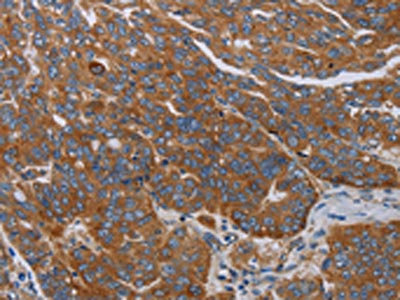
Immunohistochemical staining of human lymph node shows cytoplasmic positivity in non-germinal center cells.
Anti-PARP10 Antibody
HPA028122
ApplicationsWestern Blot, ImmunoHistoChemistry
Product group Antibodies
ReactivityHuman
TargetPARP10
Overview
- SupplierAtlas Antibodies
- Product NameAnti-PARP10 Antibody
- Delivery Days Customer4
- ApplicationsWestern Blot, ImmunoHistoChemistry
- CertificationResearch Use Only
- ClonalityPolyclonal
- ConjugateUnconjugated
- Gene ID84875
- Target namePARP10
- Target descriptionpoly(ADP-ribose) polymerase family member 10
- Target synonymsARTD10, protein mono-ADP-ribosyltransferase PARP10, ADP-ribosyltransferase diphtheria toxin-like 10, poly [ADP-ribose] polymerase 10
- HostRabbit
- IsotypeIgG
- Protein IDQ53GL7
- Protein NameProtein mono-ADP-ribosyltransferase PARP10
- Scientific DescriptionRecombinant Protein Epitope Signature Tag (PrEST) antigen sequence
- ReactivityHuman
- Storage Instruction-20°C,2°C to 8°C
- UNSPSC41116161
Datasheet
MSDS
Related products

Product group Antibodies
Anti-PARP10 Antibody Picoband(r)A08514-2-CARRIER-FREE
ApplicationsFlow Cytometry, Western Blot, ELISA
ReactivityHuman
TargetPARP10
- SizePrice
Product group Antibodies
Anti-PARP10 Antibody144-12815
ApplicationsWestern Blot
ReactivityHuman
TargetPARP10
- SizePrice
Product group Antibodies
Anti-PARP10 AntibodyA45588
ApplicationsImmunoHistoChemistry
ReactivityHuman
- SizePrice

Product group Antibodies
Anti-PARP10 AntibodyHPA052427
ApplicationsImmunoCytoChemistry
ReactivityHuman
TargetPARP10
- SizePrice
Product group Antibodies
Anti-PARP10 AntibodyHPA052427
ApplicationsImmunoCytoChemistry
ReactivityHuman
TargetPARP10
- SizePrice
Product group Antibodies

PARP10 AntibodyLS-C403409
ApplicationsELISA, ImmunoHistoChemistry
ReactivityHuman
TargetPARP10
- SizePrice
Product group Antibodies
PARP10 AntibodyCSB-PA776699
ApplicationsELISA, ImmunoHistoChemistry
ReactivityHuman
TargetPARP10
- SizePrice