


Western blot analysis of lysates from human brain, mouse brain, mouse cerebellum, rat brain tissue (from left to right), using PCGF1 Antibody (Center). A08147 was diluted at 1:1000 at each lane. A goat anti-rabbit IgG H&L (HRP) at 1:10000 dilution was used as the secondary antibody. Lysates at 20ug per lane.
Western blot analysis of lysates from human brain, mouse brain, mouse cerebellum, rat brain tissue (from left to right), using PCGF1 Antibody (Center). A08147 was diluted at 1:1000 at each lane. A goat anti-rabbit IgG H&L (HRP) at 1:10000 dilution was used as the secondary antibody. Lysates at 20ug per lane.
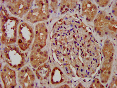
Anti-PCGF1 Antibody (Center)
A08147
ApplicationsWestern Blot
Product group Antibodies
ReactivityBovine, Human, Mouse, Rat
TargetPCGF1
Overview
- SupplierBoster Bio
- Product NameAnti-PCGF1 Antibody (Center)
- Delivery Days Customer9
- ApplicationsWestern Blot
- CertificationResearch Use Only
- ClonalityPolyclonal
- Gene ID84759
- Target namePCGF1
- Target descriptionpolycomb group ring finger 1
- Target synonyms2010002K04Rik, NSPC1, RNF3A-2, RNF68, polycomb group RING finger protein 1, RING finger protein 68, nervous system Polycomb-1
- HostRabbit
- IsotypeIgG
- Protein IDQ9BSM1
- Protein NamePolycomb group RING finger protein 1
- Scientific DescriptionBoster Bio Anti-PCGF1 Antibody (Center) (Catalog # A08147). Tested in WB application(s). This antibody reacts with Human, Mouse, Rat.
- ReactivityBovine, Human, Mouse, Rat
- Storage Instruction-20°C,2°C to 8°C
- UNSPSC12352203
Related products
Product group Antibodies
Anti-PCGF1 (Center) Antibody102-24443
ApplicationsWestern Blot
TargetPCGF1
- SizePrice
Product group Antibodies
Pcgf1 Polyclonal AntibodyCAC11417
ApplicationsImmunoFluorescence, ELISA, ImmunoHistoChemistry
TargetPCGF1
- SizePrice
Product group Antibodies
PCGF1 Polyclonal AntibodyBS-4958R
ApplicationsImmunoFluorescence, Western Blot, ELISA, ImmunoCytoChemistry, ImmunoHistoChemistry, ImmunoHistoChemistry Frozen, ImmunoHistoChemistry Paraffin
ReactivityBovine, Canine, Equine, Human, Mouse, Rabbit, Rat
- SizePrice

Product group Antibodies
Anti-PCGF1 AntibodyHPA011356
ApplicationsImmunoCytoChemistry, ImmunoHistoChemistry
ReactivityHuman
TargetPCGF1
- SizePrice
Product group Antibodies
PCGF1 AntibodyCSB-PA863114LA01HU
ApplicationsImmunoFluorescence, ELISA, ImmunoHistoChemistry
ReactivityHuman
TargetPCGF1
- SizePrice
Product group Antibodies

NSPC1 / PCGF1 AntibodyLS-C672466
ApplicationsImmunoFluorescence, ELISA, ImmunoHistoChemistry, ImmunoHistoChemistry Paraffin
ReactivityHuman
TargetPCGF1
- SizePrice