


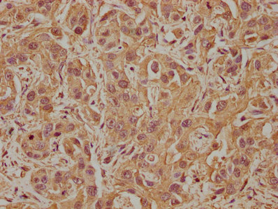
Immunohistochemical staining of human cerebral cortex shows strong cytoplasmic positivity in neuronal cells.

Immunohistochemical staining of human cerebral cortex shows strong cytoplasmic positivity in neuronal cells.
Anti-PDE8A Antibody
HPA007722
ApplicationsImmunoCytoChemistry, ImmunoHistoChemistry
Product group Antibodies
ReactivityHuman
TargetPDE8A
Overview
- SupplierAtlas Antibodies
- Product NameAnti-PDE8A Antibody
- Delivery Days Customer4
- ApplicationsImmunoCytoChemistry, ImmunoHistoChemistry
- CertificationResearch Use Only
- ClonalityPolyclonal
- ConjugateUnconjugated
- Gene ID5151
- Target namePDE8A
- Target descriptionphosphodiesterase 8A
- Target synonymsHsT19550, high affinity cAMP-specific and IBMX-insensitive 3',5'-cyclic phosphodiesterase 8A, cAMP-specific cyclic nucleotide phosphodiesterase 8A
- HostRabbit
- IsotypeIgG
- Protein IDO60658
- Protein NameHigh affinity cAMP-specific and IBMX-insensitive 3',5'-cyclic phosphodiesterase 8A
- Scientific DescriptionRecombinant Protein Epitope Signature Tag (PrEST) antigen sequence
- ReactivityHuman
- Storage Instruction-20°C,2°C to 8°C
- UNSPSC41116161
Datasheet
MSDS
Related products
Product group Antibodies
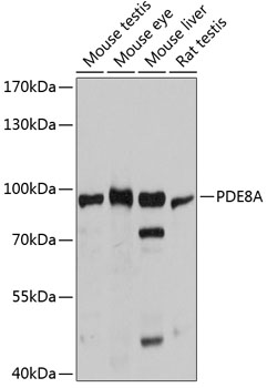
Anti-PDE8A AntibodyA80955
ApplicationsWestern Blot
ReactivityMouse, Rat
- SizePrice

Product group Antibodies
Anti-PDE8A Antibody Picoband(r)A05304-1-CARRIER-FREE
ApplicationsFlow Cytometry, Western Blot, ELISA
ReactivityHuman
TargetPDE8A
- SizePrice
Product group Antibodies
Anti-Mouse/Rat PDE8A Antibody144-12187
ApplicationsWestern Blot
ReactivityHuman, Mouse, Rat
TargetPDE8A
- SizePrice
Product group Antibodies

PDE8A AntibodyLS-C747356
ApplicationsWestern Blot
ReactivityHuman, Mouse, Rat
TargetPDE8A
- SizePrice
Product group Antibodies
PDE8A AntibodyCSB-PA525315LA01HU
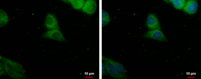
ApplicationsImmunoFluorescence, ELISA, ImmunoHistoChemistry
ReactivityHuman
TargetPDE8A
- SizePrice
Product group Antibodies
PDE8A antibodyGTX103504
ApplicationsImmunoFluorescence, ImmunoCytoChemistry
ReactivityHuman
TargetPDE8A
- SizePrice