


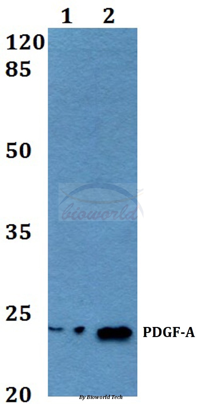
Figure 1. Western blot analysis of PDGF AA/PDGFA using anti-PDGF AA/PDGFA antibody (A02257-1). Electrophoresis was performed on a 5-20% SDS-PAGE gel at 70V (Stacking gel) / 90V (Resolving gel) for 2-3 hours. The sample well of each lane was loaded with 30 ug of sample under reducing conditions. Lane 1: human Hacat whole cell lysates, Lane 2: rat PC-12 whole cell lysates, Lane 3: mouse lung tissue lysates. After electrophoresis, proteins were transferred to a nitrocellulose membrane at 150 mA for 50-90 minutes. Blocked the membrane with 5% non-fat milk/TBS for 1.5 hour at RT. The membrane was incubated with rabbit anti-PDGF AA/PDGFA antigen affinity purified polyclonal antibody (Catalog # A02257-1) at 0.5 microg/mL overnight at 4°C, then washed with TBS-0.1%Tween 3 times with 5 minutes each and probed with a goat anti-rabbit IgG-HRP secondary antibody at a dilution of 1:5000 for 1.5 hour at RT. The signal is developed using an Enhanced Chemiluminescent detection (ECL) kit (Catalog # EK1002) with Tanon 5200 system. A specific band was detected for PDGF AA/PDGFA at approximately 24 kDa. The expected band size for PDGF AA/PDGFA is at 24 kDa.
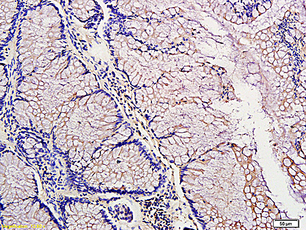
. PDGF AA/PDGFA was detected in an immunocytochemical section of SiHa cells. Enzyme antigen retrieval was performed using IHC enzyme antigen retrieval reagent (AR0022) for 15 mins. The cells were blocked with 10% goat serum. And then incubated with 5 microg/mL rabbit anti-PDGF AA/PDGFA Antibody (A02257-1) overnight at 4°C. DyLight®488 Conjugated Goat Anti-Rabbit IgG (BA1127) was used as secondary antibody at 1:100 dilution and incubated for 30 minutes at 37°C. The section was counterstained with DAPI. Visualize using a fluorescence microscope and filter sets appropriate for the label used.")
Figure 1. Western blot analysis of PDGF AA/PDGFA using anti-PDGF AA/PDGFA antibody (A02257-1). Electrophoresis was performed on a 5-20% SDS-PAGE gel at 70V (Stacking gel) / 90V (Resolving gel) for 2-3 hours. The sample well of each lane was loaded with 30 ug of sample under reducing conditions. Lane 1: human Hacat whole cell lysates, Lane 2: rat PC-12 whole cell lysates, Lane 3: mouse lung tissue lysates. After electrophoresis, proteins were transferred to a nitrocellulose membrane at 150 mA for 50-90 minutes. Blocked the membrane with 5% non-fat milk/TBS for 1.5 hour at RT. The membrane was incubated with rabbit anti-PDGF AA/PDGFA antigen affinity purified polyclonal antibody (Catalog # A02257-1) at 0.5 microg/mL overnight at 4°C, then washed with TBS-0.1%Tween 3 times with 5 minutes each and probed with a goat anti-rabbit IgG-HRP secondary antibody at a dilution of 1:5000 for 1.5 hour at RT. The signal is developed using an Enhanced Chemiluminescent detection (ECL) kit (Catalog # EK1002) with Tanon 5200 system. A specific band was detected for PDGF AA/PDGFA at approximately 24 kDa. The expected band size for PDGF AA/PDGFA is at 24 kDa.
Anti-PDGF AA/PDGFA Antibody Picoband(r)
A02257-1-CARRIER-FREE
ApplicationsImmunoFluorescence, Western Blot, ELISA, ImmunoCytoChemistry
Product group Antibodies
ReactivityHuman, Mouse, Rat
TargetPDGFA
Overview
- SupplierBoster Bio
- Product NameAnti-PDGF AA/PDGFA Antibody Picoband(r)
- Delivery Days Customer9
- ApplicationsImmunoFluorescence, Western Blot, ELISA, ImmunoCytoChemistry
- CertificationResearch Use Only
- ClonalityPolyclonal
- Concentration500 ug/ml
- Gene ID5154
- Target namePDGFA
- Target descriptionplatelet derived growth factor subunit A
- Target synonymsPDGF-A, PDGF1, platelet-derived growth factor subunit A, PDGF A-chain, PDGF subunit A, platelet-derived growth factor A-chain, platelet-derived growth factor alpha chain, platelet-derived growth factor alpha polypeptide
- HostRabbit
- IsotypeIgG
- Protein IDP04085
- Protein NamePlatelet-derived growth factor subunit A
- Scientific DescriptionBoster Bio Anti-PDGF AA/PDGFA Antibody Picoband® catalog # A02257-1. Tested in ELISA, IF, ICC, WB applications. This antibody reacts with Human, Mouse, Rat. The brand Picoband indicates this is a premium antibody that guarantees superior quality, high affinity, and strong signals with minimal background in Western blot applications. Only our best-performing antibodies are designated as Picoband, ensuring unmatched performance.
- ReactivityHuman, Mouse, Rat
- Storage Instruction-20°C,2°C to 8°C
- UNSPSC12352203
Related products
Product group Antibodies
Anti-PDGFA (N-term) Antibody102-22636
ApplicationsWestern Blot, ImmunoHistoChemistry, ImmunoHistoChemistry Paraffin
TargetPDGFA
- SizePrice
Product group Antibodies
ApplicationsWestern Blot, ImmunoHistoChemistry
ReactivityHuman, Mouse, Rat
- SizePrice
Product group Antibodies
References
PDGF-A Polyclonal AntibodyBS-0196R
ApplicationsFlow Cytometry, ImmunoFluorescence, Western Blot, ELISA, ImmunoCytoChemistry, ImmunoHistoChemistry, ImmunoHistoChemistry Frozen, ImmunoHistoChemistry Paraffin
ReactivityHuman, Mouse, Rabbit, Rat
TargetPDGFA
- SizePrice
Product group Antibodies
PDGFA AntibodyCSB-PA003723
ApplicationsWestern Blot, ELISA, ImmunoHistoChemistry
ReactivityHuman, Mouse, Rat
TargetPDGFA
- SizePrice
Product group Antibodies
ApplicationsImmunoPrecipitation, Western Blot, ImmunoCytoChemistry, ImmunoHistoChemistry
ReactivityMouse
TargetPDGFA
- SizePrice
Product group Antibodies

PDGF-AA Antibody (Preservative Free)LS-C343508
ApplicationsWestern Blot, ELISA
ReactivityHuman
TargetPDGFA
- SizePrice

Product group Antibodies
Anti-PDGFA AntibodyHPA065024
ApplicationsImmunoCytoChemistry
ReactivityHuman
TargetPDGFA
- SizePrice
Product group Antibodies
PDGFA antibodyGTX110647
ApplicationsWestern Blot
ReactivityHuman
TargetPDGFA
- SizePrice