


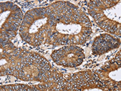
Immunohistochemical staining of human colon shows distinct positivity in plasma.

Immunohistochemical staining of human colon shows distinct positivity in plasma.
Anti-PDGFC Antibody
HPA009134
ApplicationsImmunoCytoChemistry, ImmunoHistoChemistry
Product group Antibodies
ReactivityHuman
TargetPDGFC
Overview
- SupplierAtlas Antibodies
- Product NameAnti-PDGFC Antibody
- Delivery Days Customer4
- ApplicationsImmunoCytoChemistry, ImmunoHistoChemistry
- CertificationResearch Use Only
- ClonalityPolyclonal
- ConjugateUnconjugated
- Gene ID56034
- Target namePDGFC
- Target descriptionplatelet derived growth factor C
- Target synonymsFALLOTEIN, SCDGF, platelet-derived growth factor C, PDGF-C, VEGF-E, secretory growth factor-like protein, spinal cord-derived growth factor
- HostRabbit
- IsotypeIgG
- Protein IDQ9NRA1
- Protein NamePlatelet-derived growth factor C
- Scientific DescriptionRecombinant Protein Epitope Signature Tag (PrEST) antigen sequence
- ReactivityHuman
- Storage Instruction-20°C,2°C to 8°C
- UNSPSC41116161
Datasheet
MSDS
Related products

Product group Antibodies
Anti-PDGFC Antibody Picoband(r)A03092-2-CARRIER-FREE
ApplicationsFlow Cytometry, Western Blot, ELISA
ReactivityHuman
TargetPDGFC
- SizePrice
Product group Antibodies
Anti-PDGFC AntibodyA89739
ApplicationsWestern Blot
ReactivityHuman, Mouse, Rat
- SizePrice
Product group Antibodies
PDGFC AntibodyCSB-PA108491
ApplicationsELISA, ImmunoHistoChemistry
ReactivityHuman, Mouse, Rat
TargetPDGFC
- SizePrice
Product group Antibodies

PDGF-CC AntibodyLS-C403418
ApplicationsELISA, ImmunoHistoChemistry
ReactivityHuman, Mouse, Rat
TargetPDGFC
- SizePrice
Product group Antibodies
References
PDGFC Polyclonal AntibodyBS-5775R
ApplicationsImmunoFluorescence, Western Blot, ELISA, ImmunoCytoChemistry, ImmunoHistoChemistry, ImmunoHistoChemistry Frozen, ImmunoHistoChemistry Paraffin
ReactivityBovine, Canine, Equine, Human, Mouse, Porcine, Rabbit, Rat
TargetPDGFC
- SizePrice
Product group Antibodies
ApplicationsImmunoPrecipitation, Western Blot, ImmunoCytoChemistry, ImmunoHistoChemistry
ReactivityPorcine, Rat
TargetPDGFC
- SizePrice
Product group Antibodies
PDGFC antibodyGTX51652
ApplicationsWestern Blot, ImmunoHistoChemistry, ImmunoHistoChemistry Paraffin
ReactivityHuman, Mouse
TargetPDGFC
- SizePrice
Product group Antibodies
Anti-PDGFC Antibody144-63619
ApplicationsWestern Blot
ReactivityHuman, Mouse, Rat
TargetPDGFC
- SizePrice