


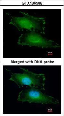
Immunofluorescent staining of human cell line PC-3 shows localization to nucleoplasm, cytosol & vesicles.

Immunofluorescent staining of human cell line PC-3 shows localization to nucleoplasm, cytosol & vesicles.
Anti-PEF1 Antibody
HPA061608
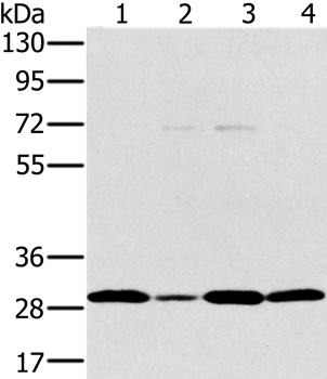
ApplicationsWestern Blot, ImmunoCytoChemistry
Product group Antibodies
ReactivityHuman
TargetPEF1
Overview
- SupplierAtlas Antibodies
- Product NameAnti-PEF1 Antibody
- Delivery Days Customer4
- ApplicationsWestern Blot, ImmunoCytoChemistry
- CertificationResearch Use Only
- ClonalityPolyclonal
- ConjugateUnconjugated
- Gene ID553115
- Target namePEF1
- Target descriptionpenta-EF-hand domain containing 1
- Target synonymsABP32, PEF1A, peflin, PEF protein with a long N-terminal hydrophobic domain, epididymis secretory sperm binding protein
- HostRabbit
- IsotypeIgG
- Protein IDQ9UBV8
- Protein NamePeflin
- Scientific DescriptionRecombinant Protein Epitope Signature Tag (PrEST) antigen sequence
- ReactivityHuman
- Storage Instruction-20°C,2°C to 8°C
- UNSPSC41116161
Datasheet
MSDS
Related products
Product group Antibodies
PEF1 AntibodyCSB-PA289634
ApplicationsWestern Blot, ELISA, ImmunoHistoChemistry
ReactivityHuman, Mouse, Rat
TargetPEF1
- SizePrice

Product group Antibodies
Anti-PEF1 Antibody Picoband(r)A09591-2-CARRIER-FREE
ApplicationsWestern Blot, ELISA, ImmunoHistoChemistry
ReactivityHuman, Mouse, Rat
TargetPEF1
- SizePrice
Product group Antibodies
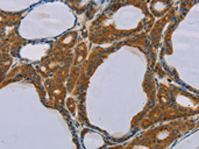
Anti-PEF1 AntibodyA38759
ApplicationsWestern Blot, ImmunoHistoChemistry
ReactivityHuman
- SizePrice
Product group Antibodies

PEF1 AntibodyLS-C753677
ApplicationsImmunoPrecipitation, Western Blot, ELISA
ReactivityHuman
TargetPEF1
- SizePrice
Product group Antibodies
PEF1 antibody [N2C3]GTX106588
ApplicationsImmunoFluorescence, Western Blot, ImmunoCytoChemistry, ImmunoHistoChemistry, ImmunoHistoChemistry Paraffin
ReactivityHuman
TargetPEF1
- SizePrice
Product group Antibodies
ApplicationsFlow Cytometry, Western Blot
ReactivityHuman, Mouse, Rat
TargetPEF1
- SizePrice
Product group Antibodies
Anti-PEF1 Antibody107-10632
ApplicationsImmunoFluorescence, Western Blot, ImmunoCytoChemistry, ImmunoHistoChemistry, ImmunoHistoChemistry Paraffin
ReactivityHuman
TargetPEF1
- SizePrice