


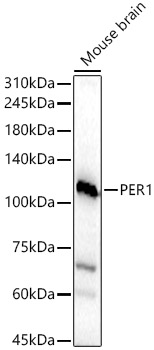
Figure 1. Western blot analysis of PER1 using anti-PER1 antibody (A00876). Electrophoresis was performed on a 5-20% SDS-PAGE gel at 70V (Stacking gel) / 90V (Resolving gel) for 2-3 hours. The sample well of each lane was loaded with 30 ug of sample under reducing conditions. Lane 1: human A549 whole cell lysates, Lane 2: human 293T whole cell lysates, Lane 3: humna A431 whole cell lysates, Lane 4: human Hela whole cell lysates, Lane 5: rat brain tissue lysates, Lane 6: rat PC-12 whole cell lysates, Lane 7: mouse brain tissue lysates, Lane 8: mouse HEPA1-6 whole cell lysates. After electrophoresis, proteins were transferred to a nitrocellulose membrane at 150 mA for 50-90 minutes. Blocked the membrane with 5% non-fat milk/TBS for 1.5 hour at RT. The membrane was incubated with rabbit anti-PER1 antigen affinity purified polyclonal antibody (Catalog # A00876) at 0.5 microg/mL overnight at 4°C, then washed with TBS-0.1%Tween 3 times with 5 minutes each and probed with a goat anti-rabbit IgG-HRP secondary antibody at a dilution of 1:5000 for 1.5 hour at RT. The signal is developed using an Enhanced Chemiluminescent detection (ECL) kit (Catalog # EK1002) with Tanon 5200 system. A specific band was detected for PER1 at approximately 200 kDa. The expected band size for PER1 is at 136 kDa.
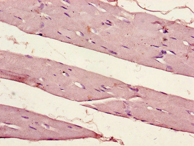
. PER1 was detected in a paraffin-embedded section of human gastric cancer tissue. Heat mediated antigen retrieval was performed in EDTA buffer (pH 8.0, epitope retrieval solution). The tissue section was blocked with 10% goat serum. The tissue section was then incubated with 2 microg/ml rabbit anti-PER1 Antibody (A00876) overnight at 4°C. Peroxidase Conjugated Goat Anti-rabbit IgG was used as secondary antibody and incubated for 30 minutes at 37°C. The tissue section was developed using HRP Conjugated Rabbit IgG Super Vision Assay Kit (Catalog # SV0002) with DAB as the chromogen.")
. PER1 was detected in an immunocytochemical section of SiHa cells. Enzyme antigen retrieval was performed using IHC enzyme antigen retrieval reagent (AR0022) for 15 mins. The cells were blocked with 10% goat serum. And then incubated with 5 microg/mL rabbit anti-PER1 Antibody (A00876) overnight at 4°C. DyLight®488 Conjugated Goat Anti-Rabbit IgG (BA1127) was used as secondary antibody at 1:500 dilution and incubated for 30 minutes at 37°C. Visualize using a fluorescence microscope and filter sets appropriate for the label used.")
Figure 1. Western blot analysis of PER1 using anti-PER1 antibody (A00876). Electrophoresis was performed on a 5-20% SDS-PAGE gel at 70V (Stacking gel) / 90V (Resolving gel) for 2-3 hours. The sample well of each lane was loaded with 30 ug of sample under reducing conditions. Lane 1: human A549 whole cell lysates, Lane 2: human 293T whole cell lysates, Lane 3: humna A431 whole cell lysates, Lane 4: human Hela whole cell lysates, Lane 5: rat brain tissue lysates, Lane 6: rat PC-12 whole cell lysates, Lane 7: mouse brain tissue lysates, Lane 8: mouse HEPA1-6 whole cell lysates. After electrophoresis, proteins were transferred to a nitrocellulose membrane at 150 mA for 50-90 minutes. Blocked the membrane with 5% non-fat milk/TBS for 1.5 hour at RT. The membrane was incubated with rabbit anti-PER1 antigen affinity purified polyclonal antibody (Catalog # A00876) at 0.5 microg/mL overnight at 4°C, then washed with TBS-0.1%Tween 3 times with 5 minutes each and probed with a goat anti-rabbit IgG-HRP secondary antibody at a dilution of 1:5000 for 1.5 hour at RT. The signal is developed using an Enhanced Chemiluminescent detection (ECL) kit (Catalog # EK1002) with Tanon 5200 system. A specific band was detected for PER1 at approximately 200 kDa. The expected band size for PER1 is at 136 kDa.
Anti-PER1 Antibody Picoband(r)
A00876-CARRIER-FREE
ApplicationsImmunoFluorescence, Western Blot, ELISA, ImmunoCytoChemistry, ImmunoHistoChemistry
Product group Antibodies
ReactivityHuman, Mouse, Rat
TargetPER1
Overview
- SupplierBoster Bio
- Product NameAnti-PER1 Antibody Picoband(r)
- Delivery Days Customer9
- ApplicationsImmunoFluorescence, Western Blot, ELISA, ImmunoCytoChemistry, ImmunoHistoChemistry
- CertificationResearch Use Only
- ClonalityPolyclonal
- Concentration500 ug/ml
- Gene ID5187
- Target namePER1
- Target descriptionperiod circadian regulator 1
- Target synonymsPER, RIGUI, hPER, period circadian protein homolog 1, Period, drosophila, homolog of, circadian clock protein PERIOD 1, circadian pacemaker protein RIGUI, hPER1, period circadian clock 1, period homolog 1
- HostRabbit
- IsotypeIgG
- Protein IDO15534
- Protein NamePeriod circadian protein homolog 1
- Scientific DescriptionBoster Bio Anti-PER1 Antibody Picoband® catalog # A00876. Tested in ELISA, IF, IHC, ICC, WB applications. This antibody reacts with Human, Mouse, Rat. The brand Picoband indicates this is a premium antibody that guarantees superior quality, high affinity, and strong signals with minimal background in Western blot applications. Only our best-performing antibodies are designated as Picoband, ensuring unmatched performance.
- ReactivityHuman, Mouse, Rat
- Storage Instruction-20°C,2°C to 8°C
- UNSPSC12352203
Related products
Product group Antibodies
PER1 AntibodyCSB-PA017786LA01HU
ApplicationsELISA, ImmunoHistoChemistry
ReactivityHuman
TargetPER1
- SizePrice
Product group Antibodies
Anti-PER1 AntibodyA16201
ApplicationsImmunoFluorescence, Western Blot, ImmunoCytoChemistry, ImmunoHistoChemistry
ReactivityHuman, Mouse, Rat
- SizePrice

Product group Antibodies
Anti-PER1 AntibodyHPA047947
ApplicationsImmunoCytoChemistry
ReactivityHuman
TargetPER1
- SizePrice
Product group Antibodies

PER1 AntibodyLS-C409984
ApplicationsWestern Blot, ImmunoHistoChemistry
ReactivityHuman, Mouse, Rat
TargetPER1
- SizePrice
Product group Antibodies
PER1 Polyclonal AntibodyBS-2350R
ApplicationsImmunoFluorescence, Western Blot, ELISA, ImmunoCytoChemistry, ImmunoHistoChemistry, ImmunoHistoChemistry Frozen, ImmunoHistoChemistry Paraffin
ReactivityBovine, Canine, Equine, Human, Mouse, Porcine, Rabbit, Rat
TargetPER1
- SizePrice
Product group Antibodies
ApplicationsImmunoPrecipitation, Western Blot, ImmunoCytoChemistry, ImmunoHistoChemistry
ReactivityMouse, Rat
TargetPER1
- SizePrice
Product group Antibodies
PER1 antibodyGTX128966
ApplicationsWestern Blot
ReactivityHuman
TargetPER1
- SizePrice
Product group Antibodies
Anti-PER1 Antibody144-08449
ApplicationsImmunoFluorescence, Western Blot, ImmunoHistoChemistry
ReactivityHuman, Mouse, Rat
TargetPER1
- SizePrice