


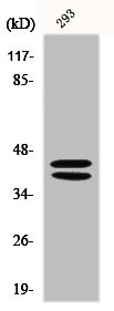
Figure 1. Western blot analysis of MAPK3 using anti-MAPK3 antibody (P00104). Electrophoresis was performed on a 5-20% SDS-PAGE gel at 70V (Stacking gel) / 90V (Resolving gel) for 2-3 hours. The sample well of each lane was loaded with 30 ug of sample under reducing conditions. Lane 1: human Hela whole cell lysates, Lane 2: human HepG2 whole cell lysates, Lane 3: human A431 whole cell lysates, Lane 4: human MCF-7 whole cell lysates, Lane 5: rat skin tissue lysates, Lane 7: mouse skiin tissue lysates. After electrophoresis, proteins were transferred to a nitrocellulose membrane at 150 mA for 50-90 minutes. Blocked the membrane with 5% non-fat milk/TBS for 1.5 hour at RT. The membrane was incubated with rabbit anti-MAPK3 antigen affinity purified monoclonal antibody (Catalog # P00104) at 1:500 overnight at 4°C, then washed with TBS-0.1%Tween 3 times with 5 minutes each and probed with a goat anti-rabbit IgG-HRP secondary antibody at a dilution of 1:500 for 1.5 hour at RT. The signal is developed using an Enhanced Chemiluminescent detection (ECL) kit (Catalog # EK1002) with Tanon 5200 system. A specific band was detected for MAPK3 at approximately 39 kDa. The expected band size for MAPK3 is at 42 kDa.
Figure 1. Western blot analysis of MAPK3 using anti-MAPK3 antibody (P00104). Electrophoresis was performed on a 5-20% SDS-PAGE gel at 70V (Stacking gel) / 90V (Resolving gel) for 2-3 hours. The sample well of each lane was loaded with 30 ug of sample under reducing conditions. Lane 1: human Hela whole cell lysates, Lane 2: human HepG2 whole cell lysates, Lane 3: human A431 whole cell lysates, Lane 4: human MCF-7 whole cell lysates, Lane 5: rat skin tissue lysates, Lane 7: mouse skiin tissue lysates. After electrophoresis, proteins were transferred to a nitrocellulose membrane at 150 mA for 50-90 minutes. Blocked the membrane with 5% non-fat milk/TBS for 1.5 hour at RT. The membrane was incubated with rabbit anti-MAPK3 antigen affinity purified monoclonal antibody (Catalog # P00104) at 1:500 overnight at 4°C, then washed with TBS-0.1%Tween 3 times with 5 minutes each and probed with a goat anti-rabbit IgG-HRP secondary antibody at a dilution of 1:500 for 1.5 hour at RT. The signal is developed using an Enhanced Chemiluminescent detection (ECL) kit (Catalog # EK1002) with Tanon 5200 system. A specific band was detected for MAPK3 at approximately 39 kDa. The expected band size for MAPK3 is at 42 kDa.
Anti-Phospho-Erk1 (T202/Y204) + Erk2 (T185/Y187) MAPK3 Rabbit Monoclonal Antibody
P00104
ApplicationsImmunoPrecipitation, Western Blot
Product group Antibodies
ReactivityHuman, Mouse, Rat
TargetMAPK1
Overview
- SupplierBoster Bio
- Product NameAnti-Phospho-Erk1 (T202/Y204) + Erk2 (T185/Y187) MAPK3 Rabbit Monoclonal Antibody
- Delivery Days Customer9
- ApplicationsImmunoPrecipitation, Western Blot
- CertificationResearch Use Only
- ClonalityMonoclonal
- Clone IDBIH-13
- Gene ID5594
- Target nameMAPK1
- Target descriptionmitogen-activated protein kinase 1
- Target synonymsERK, ERK-2, ERK2, ERT1, MAPK2, NS13, P42MAPK, PRKM1, PRKM2, p38, p40, p41, p41mapk, p42-MAPK, mitogen-activated protein kinase 1, MAP kinase 1, MAP kinase 2, MAPK 2, extracellular signal-regulated kinase 2, mitogen-activated protein kinase 2, protein tyrosine kinase ERK2
- HostRabbit
- IsotypeIgG
- Protein IDP27361
- Protein NameMitogen-activated protein kinase 3
- Scientific DescriptionBoster Bio Anti-Phospho-Erk1 (T202/Y204) + Erk2 (T185/Y187) MAPK3 Rabbit Monoclonal Antibody catalog # P00104. Tested in WB, IP applications. This antibody reacts with Human, Mouse, Rat.
- ReactivityHuman, Mouse, Rat
- Storage Instruction-20°C
- UNSPSC12352203
References
- Sun C, Xu Y, Xu G, et al. Active fractions from Jingfang Baidu Powder alleviate Klebsiella-induced Pneumonia by inhibiting TLR4/Myd88-ERK signaling pathway. J Ethnopharmacol. 2024,330:118067. doi: 10.1016/j.jep.2024.118067Read this paper
- Zhang M, Zhang Q, Zhao W, et al. The mechanism of blood coagulation induced by sodium dehydroacetate via the regulation of the mTOR/ERK pathway in rats. Toxicol Lett. 2024,392:1-11. doi: 10.1016/j.toxlet.2023.12.009Read this paper
- Zheng Y, Liu D, Guo H, et al. Paternal methamphetamine exposure induces higher sensitivity to methamphetamine in male offspring through driving ADRB1 on CaMKII-positive neurons in mPFC. Transl Psychiatry. 2023,13(1):324. doi: 10.1038/s41398-023-02624-xRead this paper
- Wei X, Chang J, Cheng Z, et al. mPFC DUSP1 mediates adolescent cocaine exposure-induced higher sensitivity to drug in adulthood. EMBO Rep. 2023,24(9):e56981. doi: 10.15252/embr.202356981Read this paper
- Chen L, Liu Z, Zhao Z, et al. Dopamine receptor 1 on CaMKII-positive neurons within claustrum mediates adolescent cocaine exposure-induced anxiety-like behaviors and electro-acupuncture therapy. Theranostics. 2023,13(10):3149-3164. doi: 10.7150/thno.83079Read this paper
- Zhang M, Li J, Ma R, et al. (2E)-1-(2,4,6-Trimethoxyphenyl)-3-(4-chlorophenyl)prop-2-en-1-one, a Chalcone Derivative, Promotes Apoptosis by Suppressing RAS-ERK and AKT/FOXO3a Pathways in Hepatocellular Carcinoma Cells. Chem Biodivers. 2023,20(7):e202300050. doi: 10.1002/cbdv.202300050Read this paper
- Wang S, Zhang Z, Liang J, et al. Identification of several inflammation-related genes based on bioinformatics and experiments. Int Immunopharmacol. 2023,121:110409. doi: 10.1016/j.intimp.2023.110409Read this paper
- Mostafa-Hedeab G, Ewaiss Hassan M, F Halawa T, et al. Epigallocatechin gallate ameliorates tetrahydrochloride-induced liver toxicity in rats via inhibition of TGFβ / p-ERK/p-Smad1/2 signaling, antioxidant, anti-inflammatory activity. Saudi Pharm J. 2022,30(9):1293-1300. doi: 10.1016/j.jsps.2022.06.021Read this paper
- Bai X, Liu X, Li S, et al. Nuciferine Inhibits TMEM16A in Dietary Adjuvant Therapy for Lung Cancer. J Agric Food Chem. 2022,70(12):3687-3696. doi: 10.1021/acs.jafc.1c08375Read this paper
- Wang YY, Wang WC, Su CW, et al. Overexpression of sprouty 1 protein in human oral squamous cell carcinogenesis. J Dent Sci. 2021,16(1):21-28. doi: 10.1016/j.jds.2020.07.013Read this paper
Datasheet
MSDS
Related products
Product group Antibodies
MAPK3/MAPK1 AntibodyCSB-PA002419
ApplicationsImmunoFluorescence, Western Blot, ELISA
ReactivityHuman, Mouse, Rat
TargetMAPK1
- SizePrice
Product group Antibodies
Anti-ERK1/2 AntibodyA29693
ApplicationsImmunoFluorescence, Western Blot, ImmunoHistoChemistry
ReactivityHuman, Mouse, Rat
- SizePrice
Product group Antibodies

Goat anti-ERK2 / MAPK1EB06592
ApplicationsImmunoFluorescence, Western Blot, ELISA
ReactivityBovine, Canine, Human, Mouse, Rat
TargetMAPK1
- SizePrice

Product group Antibodies
Anti-MAPK1-25ulHPA030069
ApplicationsWestern Blot, ImmunoHistoChemistry
ReactivityHuman
- SizePrice
Product group Antibodies

ApplicationsELISA
ReactivityHuman
TargetMAPK1
- SizePrice

Product group Antibodies
References
ApplicationsFlow Cytometry, ImmunoFluorescence, ImmunoPrecipitation, Western Blot, ImmunoCytoChemistry, ImmunoHistoChemistry
ReactivityHuman, Mouse, Rat
TargetMAPK1
- SizePrice
Product group Antibodies
MAPK1 Polyclonal AntibodyCAC14542
ApplicationsImmunoFluorescence, Western Blot, ELISA, ImmunoHistoChemistry
ReactivityRat
TargetMAPK1
- SizePrice
Product group Antibodies
References
ApplicationsFlow Cytometry, ImmunoFluorescence, Western Blot, ELISA, ImmunoCytoChemistry, ImmunoHistoChemistry, ImmunoHistoChemistry Frozen, ImmunoHistoChemistry Paraffin
ReactivityBovine, Canine, Chicken, Equine, Guinea Pig, Human, Mouse, Porcine, Rabbit, Rat
TargetMAPK1
- SizePrice